. 2006 Aug;141(4):1338-48.
doi: 10.1104/pp.106.083022. Epub 2006 Jun 30.

Induction of differentiation in the shoot apical meristem by transient overexpression of a retinoblastoma-related protein

Affiliations

Induction of differentiation in the shoot apical meristem by transient overexpression of a retinoblastoma-related protein

Joanna Wyrzykowska et al. Plant Physiol. 2006 Aug.

Abstract

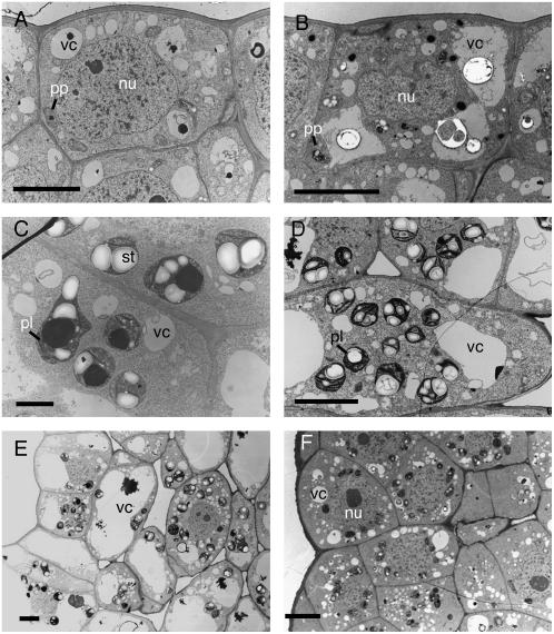
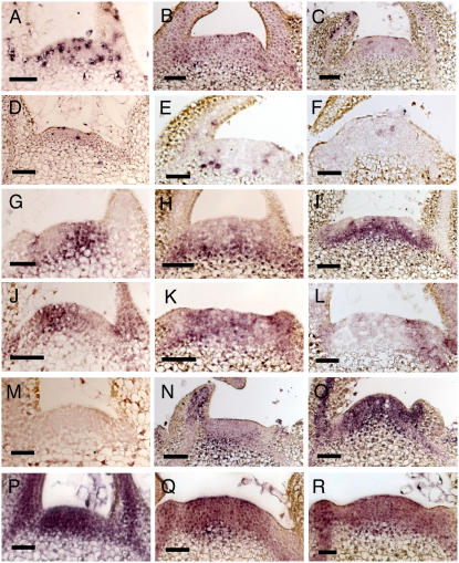
The shoot apical meristem contains cells that undergo continual growth and division to generate the building blocks for the aerial portion of the plant. As cells leave the meristem, they undergo differentiation to form specific cell types. Most notably, heterotrophic cells of the meristem rapidly gain autotrophic capability by synthesis and assembly of components of the chloroplast. At the same time, cells undergo enlargement via vacuolation. Despite significant advances in the characterization of transcriptional networks involved in meristem maintenance and leaf determination, our understanding of the actual mechanism of meristem cell differentiation remains very limited. Using a microinduction technique, we show that local, transient overexpression of a retinoblastoma-related (RBR) protein in the shoot apical meristem is sufficient to trigger cells in the meristem to undergo the initial stages of differentiation. Taken together with recent data showing that RBR protein plays a key role in restricting stem cell differentiation in the root apical meristem, our data contribute to an emerging picture of RBR proteins as a central part of the mechanism controlling meristem cell differentiation.


Figures

Figure 1.
Figure 1.
Characterization of transgenic plants showing inducible expression of the RBR gene. A, RT-PCR analysis of AtRBR expression in leaf discs from independent lines of tobacco transformed with the Tet∷RBR construct. Lines were either induced (+) or not induced (−) with AhTet. M, Molecular size marker. B, Western-blot analysis of protein extracted from tobacco lines transformed with the Tet∷RBR construct and either induced (+) or not induced (−) with AhTet for 4 h. AtWT, Extract from wild-type Arabidopsis; 35S∷CycD, extract from transgenic Arabidopsis engineered to overexpress a cyclinD. These plants have been previously shown to contain elevated levels of the AtRBR protein (Dewitte et al., 2003). Tet∷GUS, Extract from transgenic tobacco plants engineered to inducibly express the GUS reporter gene after treatment with AhTet. Arrowhead indicates migration of a 100-kD marker protein. The bottom image shows amidoblack staining of the blot to ensure approximate equal loading of total protein in all lanes. C, Real-time PCR analysis of NtRBR (white columns) and AtRBR (shaded columns) at different time points in Tet∷RBR apices after induction of AtRBR expression. Expression of each transcript is relative to that of an endogenous actin mRNA (as described in “Materials and Methods”), and this ratio was defined as equal to 1 for the zero time point for each RBR gene. Log data derived from the analysis of three independent biological replicates, with each sample analyzed in triplicate, are shown. D, In situ hybridization analysis of meristems from Tet∷RBR plants hybridized with antisense probes for either NtRBR or AtRBR (as indicated) and at time points 0, 2, and 24 h after AhTet induction (as indicated). Longitudinal sections through the meristems are shown and signal is visible as dark staining. Bar = 50 μm.
Figure 2.
Figure 2.
Microinduction of RBR gene expression in the SAM leads to repression of growth. A and B, Apices from Tet∷RBR plants showing total repression of growth after microinduction. C and D, Apices from Tet∷RBR plants showing intermediate growth retardation after microinduction. E, Normal growth of apices from Tet∷GUS plants induced with AhTet. F, Normal growth of an apex from a Tet∷RBR plant after mock induction. Plant growth was recorded 5 weeks after microinduction. Bar in A and B = 2 mm.
Figure 3.
Figure 3.
Induction of RBR gene expression leads to altered meristem histology. A, Longitudinal section of a control apex showing the meristem dome. B, Detail of the outer cell layers from a control meristem. C, As in A, but from a Tet∷RBR meristem in which RBR gene expression has been induced by microinduction 24 h previously (arrowhead). D, Detail from the section in C showing the enlarged cells (arrowhead) observed after microinduction of RBR genes. E, As in A, but from a Tet∷RBR meristem in which RBR gene expression has been induced by microinduction 72 h previously. F, Section to show the histology of the central region of a meristem treated as in E. G, As in A, but from a Tet∷RBR meristem in which RBR expression has been induced by microinduction 3 weeks previously. H, Section to show the histology of the central region of a meristem treated as in G. I, Scanning electron micrograph of a tobacco apex to show approximate orientation (black line) of the sections shown in A to H. Primordia numbers are given as P1 and P2. Bars in A, C, E, F, G, and H = 40 μm; bars in B and D = 20 μm.
Figure 4.
Figure 4.
Histology of primordia from apices in which RBR gene expression has been locally induced. A, Longitudinal section through a primordium showing abnormal histology (retarded vacuolation and enlargement) on the induced adaxial (Ad) face of the organ compared to the abaxial (Ab) face in which a normal progression of leaf cell differentiation is occurring. B, Longitudinal section through the adaxial face of a primordium from an induced Tet∷RBR apex. C, As in B, but from a control mock-induced apex. Bar in A = 200 μm; bar in B and C = 40 μm.
Figure 5.
Figure 5.
Electron microscopy reveals promotion of differentiation in meristem cells after microinduction of RBR gene expression. A, Cell from the outer layer of a control meristem. B, Cell from the outer layer of a Tet∷RBR meristem in which RBR gene expression has been induced 24 h previously. C, Plastid differentiation in a tunica cell of a Tet∷RBR plant in which RBR gene expression has been induced 24 h previously. D, Plastid differentiation in a primordium from a control apex. E, Adaxial cell differentiation from a control leaf primordium. F, Adaxial cells from a primordium of a Tet∷RBR apex has been microinduced to express RBR 72 h previously. nu, Nucleus; pl, plastid; pp, proplastid; st, starch granule; vc, vacuole. Bars in A, B, D, E, and F = 5 μm; bar in C = 1 μm.
Figure 6.
Figure 6.
Analysis of marker gene expression in meristems microinduced to overexpress RBR gene expression. A, In situ hybridization with an antisense probe for histone H4 against a longitudinal section of an apex from a Tet∷RBR plant without AhTet induction. B, As in A, but 24 h after microinduction. C, As in A, but 72 h after microinduction. D, As in A, but hybridized with an antisense probe for NtCYCB1. E, As in D, but 24 h after microinduction. F, As in D, but 72 h after microinduction. G, As in A, but hybridized with an antisense probe against NTH15. H, As in G, but 24 h after microinduction. I, As in G, but 72 h after microinduction. J, As in A, but hybridized with an antisense probe against NTPHAN. K, As in J, but 24 h after microinduction. L, As in J, but 72 h after microinduction. M, As in A, but hybridized with an antisense probe against RBCS. N, As in M, but 24 h after microinduction. O, As in M, but 72 h after microinduction. P, As in A, but hybridized with an antisense probe against NteIF4A. Q, As in P, but 24 h after microinduction. R, As in P, but 72 h after microinduction. Bars = 50 μm.

Similar articles

Cited by

References

    1. Ach RA, Durfee T, Miller AB, Taranto P, Hanley-Bowdoin L, Zambryski PC, Gruissem W (1997) RRB1 and RRB2 encode maize retinoblastoma-related proteins that interact with a plant D-type cyclin and geminivirus replication protein. Mol Cell Biol 17: 5077–5086 - PMC - PubMed
    1. Byrne ME (2005) Networks in leaf development. Curr Opin Plant Biol 8: 59–66 - PubMed
    1. Dewitte W, Riou-Khamlichi C, Scofield S, Healy JMS, Jacqmard A, Kilby NJ, Murray JA (2003) Altered cell cycle distribution, hyperplasia, and inhibited differentiation in Arabidopsis caused by the D-type cyclin CYCD3. Plant Cell 15: 79–92 - PMC - PubMed
    1. Dimova DK, Stevaux O, Frolov MV, Dyson NJ (2003) Cell cycle-dependent and cell cycle-independent control of transcription by the Drosophila E2F/RB pathway. Genes Dev 17: 2308–2320 - PMC - PubMed
    1. Donnelly PM, Bonetta D, Tsukaya H, Dengler RE, Dengler NG (1999) Cell cycling and cell enlargement in developing leaves of Arabidopsis. Dev Biol 215: 407–419 - PubMed

Publication types

MeSH terms