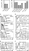
Naive CD4 T cells constitutively express CD40L and augment autoreactive B cell survival
- PMID: 16815973
- PMCID: PMC1484418
- DOI: 10.1073/pnas.0601539103
Naive CD4 T cells constitutively express CD40L and augment autoreactive B cell survival
Abstract
Chronic engagement of the B cell receptor by soluble autoantigen leads to reduced B cell survival. Using the Ig and hen egg lysozyme double transgenic mouse model, we demonstrate that the survival of soluble autoantigen-engaged B cells is further reduced in mice lacking CD4 T cells or deficient in CD40. Mixed bone marrow chimera experiments reveal that, under homeostatic conditions, the CD40L-CD40 pathway can augment autoreactive B cell survival in a non-cell-autonomous manner. Naive CD4 T cells are shown to constitutively express CD40L mRNA and protein, although cell surface CD40L abundance is low because of engagement with CD40 on other cells. These observations indicate that the CD40L-CD40 pathway can augment the survival of autoantigen-engaged B cells in the absence of T cell activation. We propose that constitutive CD40L expression by naive CD4 T cells influences the composition of the B cell repertoire and may also affect the homeostasis of other cell types such as regulatory T cells in lymphoid organs.
Conflict of interest statement
Conflict of interest statement: No conflicts declared.
Figures


Similar articles
-
Constitutive CD40L expression on B cells prematurely terminates germinal center response and leads to augmented plasma cell production in T cell areas.J Immunol. 2010 Jul 1;185(1):220-30. doi: 10.4049/jimmunol.0901689. Epub 2010 May 26. J Immunol. 2010. PMID: 20505142 Free PMC article.
-
Increased T cell autoreactivity in the absence of CD40-CD40 ligand interactions: a role of CD40 in regulatory T cell development.J Immunol. 2001 Jan 1;166(1):353-60. doi: 10.4049/jimmunol.166.1.353. J Immunol. 2001. PMID: 11123312
-
Murine Fibroblastic Reticular Cells From Lymph Node Interact With CD4+ T Cells Through CD40-CD40L.Transplantation. 2015 Aug;99(8):1561-7. doi: 10.1097/TP.0000000000000710. Transplantation. 2015. PMID: 25856408 Free PMC article.
-
The role of CD40 ligand in costimulation and T-cell activation.Immunol Rev. 1996 Oct;153:85-106. doi: 10.1111/j.1600-065x.1996.tb00921.x. Immunol Rev. 1996. PMID: 9010720 Review.
-
Regulation of CD40 ligand expression in systemic lupus erythematosus.Curr Opin Rheumatol. 2001 Sep;13(5):361-9. doi: 10.1097/00002281-200109000-00004. Curr Opin Rheumatol. 2001. PMID: 11604589 Review.
Cited by
-
Characterization of the Bronchoalveolar Lavage Fluid by Single Cell Gene Expression Analysis in Healthy Dogs: A Promising Technique.Front Immunol. 2020 Jul 30;11:1707. doi: 10.3389/fimmu.2020.01707. eCollection 2020. Front Immunol. 2020. PMID: 32849601 Free PMC article.
-
A combination of cellular biomarkers predicts failure to respond to rituximab in rheumatoid arthritis: a 24-week observational study.Arthritis Res Ther. 2016 Aug 24;18(1):190. doi: 10.1186/s13075-016-1091-1. Arthritis Res Ther. 2016. PMID: 27558631 Free PMC article.
-
Extracellular Vesicles Mediate B Cell Immune Response and Are a Potential Target for Cancer Therapy.Cells. 2020 Jun 22;9(6):1518. doi: 10.3390/cells9061518. Cells. 2020. PMID: 32580358 Free PMC article. Review.
-
Human CD14hi monocytes and myeloid dendritic cells provide a cell contact-dependent costimulatory signal for early CD40 ligand expression.Blood. 2011 Feb 3;117(5):1585-94. doi: 10.1182/blood-2008-01-130252. Epub 2010 Jul 15. Blood. 2011. PMID: 20634374 Free PMC article.
-
CD40 signaling to the rescue: A CD8 exhaustion perspective in chronic infectious diseases.Crit Rev Immunol. 2013;33(4):361-78. doi: 10.1615/critrevimmunol.2013007444. Crit Rev Immunol. 2013. PMID: 23971530 Free PMC article.
References
-
- Goodnow C. C., Sprent J., Fazekas de St Groth B., Vinuesa C. G. Nature. 2005;435:590–597. - PubMed
-
- Wardemann H., Yurasov S., Schaefer A., Young J. W., Meffre E., Nussenzweig M. C. Science. 2003;301:1374–1377. - PubMed
-
- Cyster J. G., Goodnow C. C. Immunity. 1995;3:691–701. - PubMed
-
- Lesley R., Xu Y., Kalled S. L., Hess D. M., Schwab S. R., Shu H. B., Cyster J. G. Immunity. 2004;20:441–453. - PubMed
Publication types
MeSH terms
Substances
Grants and funding
LinkOut - more resources
Full Text Sources
Research Materials

