. 2006 Jul 15;20(14):1933-45.
doi: 10.1101/gad.1411206. Epub 2006 Jun 30.

The role of microtubule actin cross-linking factor 1 (MACF1) in the Wnt signaling pathway

Affiliations

The role of microtubule actin cross-linking factor 1 (MACF1) in the Wnt signaling pathway

Hui-Jye Chen et al. Genes Dev. .

Abstract

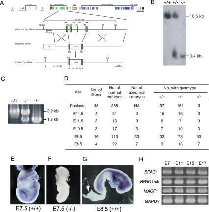
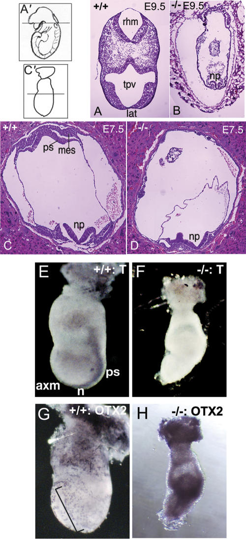
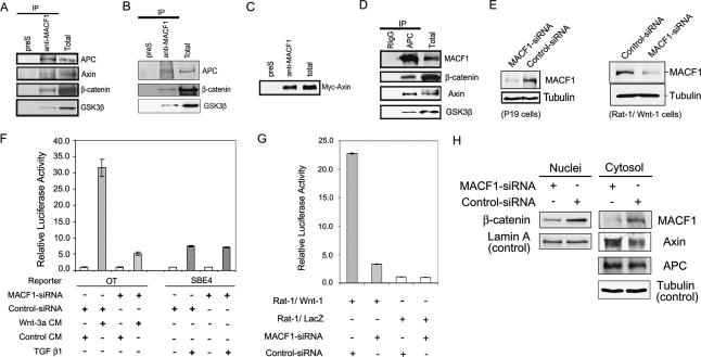
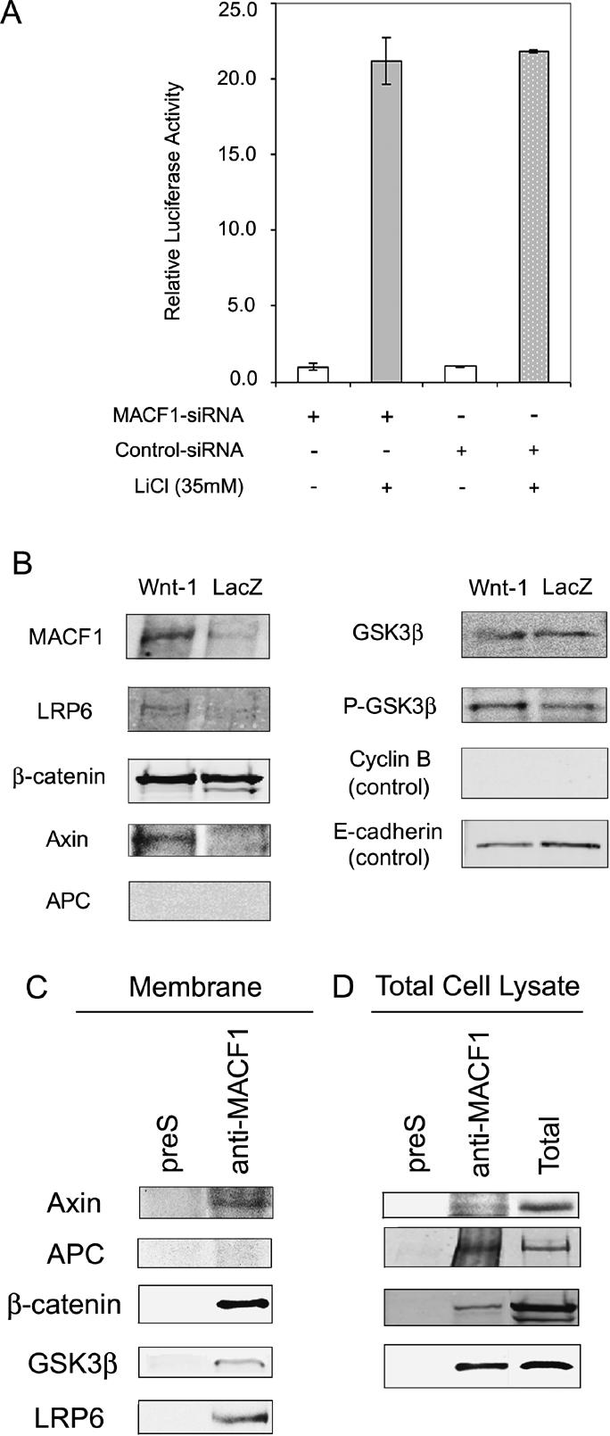
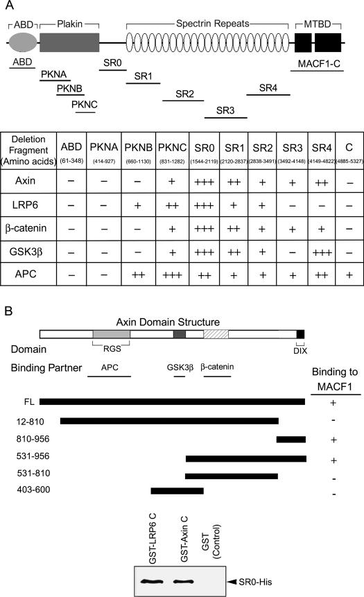
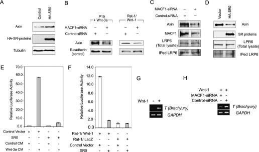
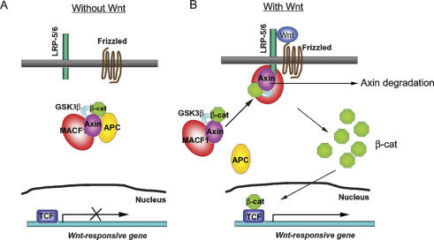
MACF1 (microtubule actin cross-linking factor 1) is a multidomain protein that can associate with microfilaments and microtubules. We found that MACF1 was highly expressed in neuronal tissues and the foregut of embryonic day 8.5 (E8.5) embryos and the head fold and primitive streak of E7.5 embryos. MACF1(-/-) mice died at the gastrulation stage and displayed developmental retardation at E7.5 with defects in the formation of the primitive streak, node, and mesoderm. This phenotype was similar to Wnt-3(-/-) and LRP5/6 double-knockout embryos. In the absence of Wnt, MACF1 associated with a complex that contained Axin, beta-catenin, GSK3beta, and APC. Upon Wnt stimulation, MACF1 appeared to be involved in the translocation and subsequent binding of the Axin complex to LRP6 at the cell membrane. Reduction of MACF1 with small interfering RNA decreased the amount of beta-catenin in the nucleus, and led to an inhibition of Wnt-induced TCF/beta-catenin-dependent transcriptional activation. Similar results were obtained with a dominant-negative MACF1 construct that contained the Axin-binding region. Reduction of MACF1 in Wnt-1-expressing P19 cells resulted in decreased T (Brachyury) gene expression, a DNA-binding transcription factor that is a direct target of Wnt/beta-catenin signaling and required for mesoderm formation. These results suggest a new role of MACF1 in the Wnt signaling pathway.


Figures

Figure 1.
Figure 1.
Targeted disruption of the mouse MACF1 gene. (A) Genomic organization and partial restriction maps of the mouse MACF1 locus, targeting vector, and targeted locus. The domains are marked above the construct. (Xb and Xh) XbaI and XhoI restriction sites; (neo) a cassette for neomycin phosphotransferase; (arrows) genotyping PCR primers; the position of the Southern blot probe is shown. (B) Southern blot analysis of E10.5 mice. The wild-type and mutant alleles generated 10.5-kb and 3.4-kb XbaI fragments, respectively. (C) PCR genotyping of E10.5 embryos. Products from wild-type animals and mutants are 3.0 kb and 1.8 kb, respectively. (+/+) Wild-type mouse; (+/−) heterozygous mouse; (−/−) homozygous-null mouse. (D) Genotypes of progeny from heterozygous matings. The pooled data from several litters are shown. (NA) Not available. (E–G) Whole-mount immunohistochemistry with anti-MACF1 antibody. (E) In the wild-type E7.5 embryo, MACF1 was expressed in the neural fold and primitive streak. (F) No MACF1 staining was observed in MACF1-null embryos. (G) In the wild-type E8.5 embryo, MACF1 protein was widely expressed, except in the allantois (arrow). (H) RT–PCR analyses of MACF1 and BPAG1 at E7, E11, E15, and E17. (BPAG1) All BPAG1 isoforms; (BPAG1a/b) BPAG1a and BPAG1b isoforms; (GAPDH) glyceraldehyde-3-phosphate dehydrogenase, loading control.
Figure 2.
Figure 2.
Histological and tissue-specific marker analyses of MACF1−/− embryos. (A–D) H&E staining of transverse cryosections of E9.5 wild-type (A) and mutant (B) embryos, and E7.5 wild-type (C) and mutant (D) embryos. (np) Neural plate; (rhm) rhombomere; (tpv) telencephalic vesicle; (lat) lamina terminalis; (ps) primitive streak; and (mes) mesoderm. A′ and C′ show the location of the sectioning site of E9.5 and E7.5 embryos, respectively. (E–H) Whole-mount in situ immunohistochemistry analyses of E7.5 wild-type (E,G) and MACF1 mutant (F,H) embryos. The embryos are shown with the anterior facing the left. T was expressed in the primitive streak (ps), node (n), and axial mesoderm (axm) of the control embryo (E); these structures were absent in the mutant embryo and resulted in no T staining (F). OTX2 is expressed in the anterior neuroectoderm of control embryo (bracket; G) and mutant embryo (H).
Figure 3.
Figure 3.
MACF1 in the Wnt/β-catenin signaling pathway. (A,B) Cell lysates from PC12 cells (A) and Rat-1/LacZ cells (B) were immunoprecipitated (IP) by anti-MACF1 antibody, and the blots were probed with anti-APC, anti-Axin, anti-β-catenin, and anti-GSK3β antibodies. (preS) Immunoprecipitation control by preimmune serum; (Total) total lysate. (C) Cell lysates from c-myc-Axin-transfected PC12 cells were immunoprecipitated by anti-MACF1 antibody, and the blot was probed with anti-c-Myc antibody. (D) Cell lysates from PC12 cells were immunoprecipitated by anti-APC antibody, and the blots were probed for MACF1, β-catenin, Axin, and GSK3β. (RigG) Immunoprecipitation control by rabbit IgG. (E) Knockdown of MACF1 protein by siRNA in Wnt-1-transfected-P19 cells (left) and Rat-1/Wnt-1 cells (right). Cell lysates were subjected to Western blots with anti-MACF1 antibody (MACF1) and anti-β-tubulin antibody (Tubulin). (F) P19 cells were transfected with MACF1 siRNA or control vector and reporter constructs pGL3-OT (OT) or pSBE4-luc (SBE4), and then treated with Wnt-3a-conditioned medium, control medium, or TGFβ 1 (200 pM). The lysates were subjected to dual luciferase assays. Each data point was done in triplicate. (G) Rat-1/Wnt-1 or Rat-1/LacZ cells were transfected with MACF1 siRNA vector or control vector, and reporter constructs. Assay conditions were the same as in F. (H) Rat-1/Wnt-1 cells were transfected with MACF1 siRNA or control vector, and nuclear (Nuclei) and cytosolic (Cytosol) fractions were isolated. The cytosolic fraction was probed for MACF1, Axin, and APC, while the nuclear fraction was probed for β-catenin. β-tubulin (Tubulin) and Lamin A were used as loading controls for cytosol and nuclear fractions, respectively.
Figure 4.
Figure 4.
MACF1 acts upstream of GSK3β and associates with the Axin complex at the cell membrane upon Wnt stimulation. (A) TCF/β-catenin transcriptional activity of MACF1 knockdown P19 cells after LiCl treatment to inhibit GSK3β activity. Cell lysates were subjected to dual luciferase assays. LiCl treatment is in gray, while no treatment is in white. (B) Membrane fractions were isolated from Rat-1/Wnt-1 or Rat-1/LacZ cells, and the blots were probed for MACF1, LRP6, β-catenin, Axin, APC, GSK3β, and phosphorylated GSK3β (P-GSK3β). Cytosolic cyclin B and membranous E-cadherin served as control. (C) The membrane fraction was isolated from Rat-1/Wnt-1 cells and immunoprecipitated by anti-MACF1 antibody, and the blots were probed for indicated proteins. (preS) Immunoprecipitation control by preimmune serum. Note that all the Axin complex components, except APC, were immunoprecipitated by anti-MACF1. (D) Total cell lysates from Rat-1/Wnt-1 cells were immunoprecipitated by anti-MACF1 antibody, and the blots were probed for indicated proteins. The blots of the total cell lysates (Total) are shown as comparison.
Figure 5.
Figure 5.
Mapping the interactions between MACF1 and Wnt signaling proteins. (A) Identification of MACF1 domains that interact with Wnt signaling proteins. The top panel shows the domain organization of MACF1. The relative positions of the truncated constructs are indicated. Interactions between MACF1 and Wnt signaling proteins were determined by coimmunoprecipitation experiments. The degree of binding is symbolized by the number of + signs; (−) no binding. (B) Dissection of the interactions between Axin and MACF1. The diagram shows the domain structure of Axin and the location of the binding regions for other proteins. + and − indicate the ability of these constructs to coimmunoprecipitate with anti-MACF1. (Bottom panel) MACF1 deletion fragment SR0 binds directly to LRP6C and Axin C. GST-pull-down assays were performed by incubating SR0-his (purified SR0 fragment with a C-terminal 6× his tag) with GST-LRP6C (the intracellular domain of LRP6 fused with GST protein; purified), GST-Axin C (C terminus of Axin fused with GST; purified), or GST proteins alone (GST; control). The blot was probed with anti-his antibody.
Figure 6.
Figure 6.
Involvement of MACF1 in Axin translocation to the cell membrane and reduction of T (Brachyury) gene expression in MACF1 knockdown P19 cells transfected with Wnt-1. (A) Overexpression of MACF1 deletion fragment SR0 resulted in an increase of Axin stability. Rat-1/Wnt-1 cells were transfected with HA-SR0 or control vectors (Control), lysed, and processed for Western blotting using anti-Axin (Axin), anti-HA (HA-SR-proteins), and anti-tubulin (Tubulin) antibodies. (B) Knockdown of MACF1 decreased the amount of Axin at the cell membrane. Rat-1/Wnt-1 cells and Wnt-3a-treated P19 cells were transfected with MACF1 siRNA vector or control vector. Membrane fractions were isolated, and the blot was probed for Axin. E-cadherin served as a control for membrane proteins. (C) Knockdown of MACF1 decreased the binding of Axin to LRP6. Rat-1/Wnt-1 cells were transfected with MACF1 siRNA or control vectors. Cell lysates were immunoprecipitated by anti-LRP6 antibody, and the blot was probed with anti-Axin (top panel) and anti-LRP6 (IPed LRP6; bottom panel) antibodies. Western blotting of the total cell lysate with anti-LRP6 antibody (LRP6; lower middle panel) and knockdown of MACF1 is shown by anti-MACF1 antibody (upper middle panel). (D) Overexpression of MACF1 deletion fragment SR0 reduced the binding of Axin to LRP6. Rat-1/Wnt-1 cells were transfected with MACF1 deletion cDNA clone HA-SR0 or control vector. Cell lysate was immunoprecipitated by anti-LRP6 antibody, and the blot was probed with anti-Axin (top panel) and anti-LRP6 (IPed LRP6; bottom panel) antibodies. Expression of SR and endogenous LRP6 proteins was shown in the blots from total cell lysate probed with anti-HA (SR proteins; upper middle panel) and anti-LRP6 (lower middle panel) antibodies. (E) Reduction of TCF/β-catenin transcriptional activity in Wnt-3a-treated P19 cells expressing SR0 proteins. P19 cells were transfected with reporter constructs, and HA-SR0 or control constructs, then treated with Wnt-3a-conditioned medium (Wnt-3a CM; in gray) or control medium (Control CM; in white). Cell lysis, dual luciferase assay, and data processing were as described in Figure 3F. (F) Reduction of TCF/β-catenin transcriptional activity in Rat-1/Wnt-1 cells expressing SR0 proteins. Rat-1/Wnt-1 cells were transfected with reporter constructs, HA-SR0, or control constructs, and assayed for luciferase activity. (Control) White bar; (SR0) gray bar. (G) T (Brachyury) gene expression could be induced by expressing Wnt-1 in P19 cells. P19 cells were transfected with Wnt-1, and T (Brachyury) gene expression was assessed by RT–PCR. GAPDH served as an internal control. (H) T (Brachyury) gene expression was decreased in MACF1-knocked-down P19 cells. P19 cells were transfected with MACF1-siRNA vector and Wnt-1 or control siRNA vector and Wnt-1 (treatment symbolized by +), then T (Brachyury) gene expression was assessed by RT–PCR as described in the Materials and Methods. GAPDH served as an internal control.
Figure 7.
Figure 7.
A model for the involvement of MACF1 in the Wnt/β-catenin signaling pathway. (A) Without Wnt, MACF1 (red) binds to a protein complex containing Axin (purple), APC (yellow), β-catenin (green), and GSK3β (light blue). There is no expression of the TCF (blue) controlled genes due to the degradation of β-catenin after phosphorylation by GSK3β. (B) Wnt activates its receptor frizzled and its coreceptor LRP-5/6. MACF1 is involved in the translocation of the complex containing Axin, β-catenin, and GSK3β but not APC from the cytosol to the cell membrane, where Axin and MACF1 bind to LRP-5/6. Subsequently, GSK3β is inactivated by phosphorylation, Axin is degraded, and β-catenin is released and enters the nucleus, where it can activate the Wnt-responsive genes.

References

    1. Arnold S.J., Stappert J., Bauer A., Kispert A., Herrmann B.G., Kemler R. Brachyury is a target gene of the Wnt/β-catenin signaling pathway. Mech. Dev. 2000;91:249–258. - PubMed
    1. Beddington R.S., Robertson E.J. Axis development and early asymmetry in mammals. Cell. 1999;96:195–209. - PubMed
    1. Bosher J.M., Hahn B.S., Legouis R., Sookhareea S., Weimer R.M., Gansmuller A., Chisholm A.D., Rose A.M., Bessereau J.L., Labouesse M. The Caenorhabditis elegans vab-10 spectraplakin isoforms protect the epidermis against internal and external forces. J. Cell Biol. 2003;161:757–768. - PMC - PubMed
    1. Chen H.J., Hwong C.L., Wang C.H., Hwang J. Degradation of DNA topoisomerase I by a novel trypsin-like serine protease in proliferating human T lymphocytes. J. Biol. Chem. 2000;275:13109–13117. - PubMed
    1. Cliffe A., Hamada F., Bienz M. A role of Dishevelled in relocating Axin to the plasma membrane during wingless signaling. Curr. Biol. 2003;13:960–966. - PubMed

Publication types

MeSH terms