Review
. 2006 Mar;5(1):41-60.
doi: 10.1177/1534582306289043.

What is the functional significance of chronic stress-induced CA3 dendritic retraction within the hippocampus?

Affiliations
Review

What is the functional significance of chronic stress-induced CA3 dendritic retraction within the hippocampus?

Cheryl D Conrad. Behav Cogn Neurosci Rev. 2006 Mar.

Abstract

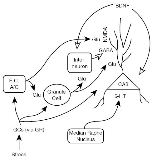
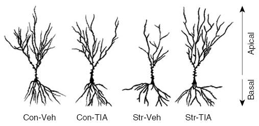
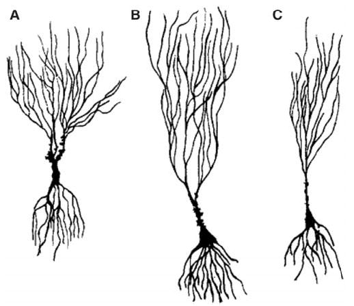
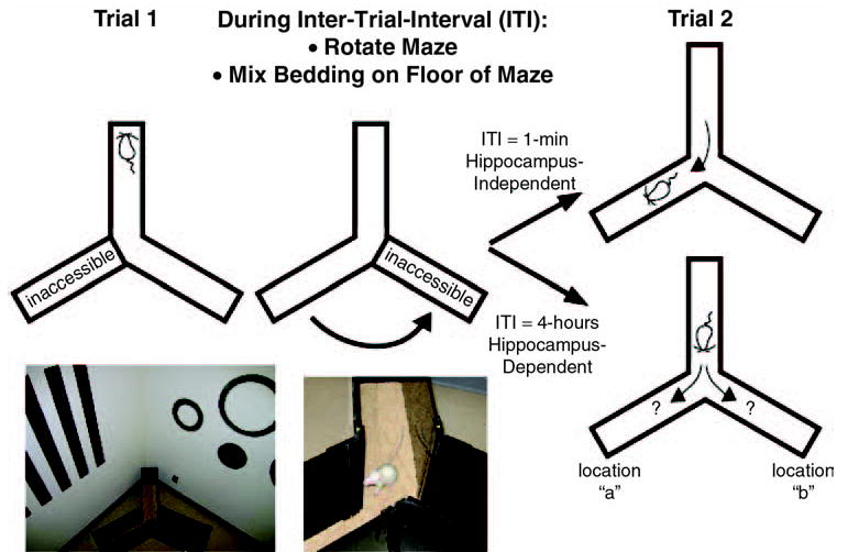
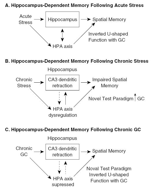
Chronic stress produces consistent and reversible changes within the dendritic arbors of CA3 hippocampal neurons, characterized by decreased dendritic length and reduced branch number. This chronic stress-induced dendritic retraction has traditionally corresponded to hippocampus-dependent spatial memory deficits. However, anomalous findings have raised doubts as to whether a CA3 dendritic retraction is sufficient to compromise hippocampal function. The purpose of this review is to outline the mechanism underlying chronic stress-induced CA3 dendritic retraction and to explain why CA3 dendritic retraction has been thought to mediate spatial memory. The anomalous findings provide support for a modified hypothesis, in which chronic stress is proposed to induce CA3 dendritic retraction, which then disrupts hypothalamic-pituitary-adrenal axis activity, leading to dysregulated glucocorticoid release. The combination of hippocampal CA3 dendritic retraction and elevated glucocorticoid release contributes to impaired spatial memory. These findings are presented in the context of clinical conditions associated with elevated glucocorticoids.


Figures

Figure 1
Figure 1. Mechanism of CA3 Dendritic Retraction Following Chronic Stress
NOTE: Repeated glucocorticoid (GC) elevations from chronic stress directly influence the CA3 pyramidal cells and CA3 afferents (dentate gyrus granule cells, commissural/associational fibers [C/A], entorhinal cortex [E.C.]) because all of these cells express receptors for GCs. The glucocorticoid receptor (GR) most likely mediates dendritic retraction in rodents, but the mineralocorticoid receptor (MR) probably plays a role in primates. Excess glutamate (Glu via N-methyl-D-aspartate [NMDA] receptor) and serotonin (5-HT) as well as altered inhibitory tone from interneurons and gamma-aminobutyric acid (GABA) modulate CA3 dendritic retraction. Reduced levels of brain-derived neurotrophic factor (BDNF), which is retrogradely transported to CA3 neurons, may permit CA3 dendritic remodeling. Solid arrows = enhanced tone permits CA3 dendritic retraction; open arrows = reduced tone permits CA3 dendritic retraction.
Figure 2
Figure 2. Camera Lucida Drawings of Hippocampal CA3 Pyramidal Cells
NOTE: Chronic restraint stress for 6 hours/day for 21 days decreased the complexity of the CA3 apical dendritic tree (Str-Veh). Daily tianeptine injections (10 mg/kg, intraperitoneally) prior to each daily restraint session prevented the CA3 apical dendritic retraction (Str-TIA) and produced dendritic arbors that were indistinguishable from controls injected with vehicle (Con-Veh) or tianeptine (Con-TIA). The CA3 basal dendritic arbors were unaffected by these manipulations. SOURCE: Reprinted with permission from Conrad, Magariños, LeDoux, and McEwen (1999).
Figure 3
Figure 3. Camera Lucida Drawings of Complex Superior Cervical Cells
NOTE: The cells are arranged from left to right according to total dendritic length. The estimate of the number of inputs received by each cell is indicated beside the axon (which extends much farther than shown). The geometrically simpler cells (A) receive fewer inputs than more complex neurons (B). SOURCE: Reprinted excerpt with permission from Purves and Lichtman (1985), the American Association for the Advancement of Science.
Figure 4
Figure 4. Camera Lucida Drawings of Pyramidal Neurons From the Motor Cortex
NOTE: These are the same kind of neuron, represented from different species. The pyramidal neurons from the motor cortices of different mammals are drawn to the same scale. Enhanced morphological complexity, as observed in dendritic arbors, is proposed to contribute to higher cognitive function. SOURCE: With permission from Rosenzweig, Breedlove, and Watson (2005).
Figure 5
Figure 5. Camera Lucida Drawings of Hippocampal CA3 Neurons From Ground Squirrels
NOTE: Short (A) and long-shafted neurons (B, C) found in ground squirrels. The long-shafted neurons showed the greatest structural reactivity to hibernation, with the CA3 neurons of awakened ground squirrels (B) showing greater structural complexity than those of ground squirrels in hibernation (C). This observation that the long-shafted neurons have a greater response to remodeling than short-shafted neurons do is similar to observations of differential responsivity of CA3 neuronal subtypes following chronic stress in female rats (McLaughlin, Baran, Wright, & Conrad, 2005). SOURCE: Reprinted from Popov, Bocharova, and Bragin (1992), with permission from Elsevier.
Figure 6
Figure 6. Schematic and Principle of the Y-Maze
NOTE: A rat is placed in one arm of a symmetrical Y-maze and allowed to explore two arms whereas the third arm is inaccessible (blocked with the same Plexiglas material as the Y-maze). After 15 minutes of exploration, the rat is returned to its home cage so that the Y-maze can be rotated and the bedding on the floor of the maze mixed to reduce the potential use of intramaze cues. After the designated intertrial interval (ITI), the rat is reintroduced to the same start location and allowed to explore all three arms. At 1-minute ITI, rats with or without a compromised hippocampus readily explore the arm in the previously unexplored location. In contrast, at 4-hour ITI, rats with a compromised hippocampus enter the arms of the unexplored and explored locations similarly. Not shown are the salient cues located outside the Y-maze that help with navigation, and these include posters, shelving, and laboratory equipment. Regarding Trial 2, please note that the previously inaccessible arm is now located in a previously explored room location (indicated as “b”), and the previously inaccessible room location is now occupied by a previously explored arm (indicated as “a”). If intramaze (hippocampus-independent) cues guide exploration of the arms, then rats are expected to explore location “b” more than location “a.” However, preference for location “b” is rarely observed because rats either prefer the novel location “a” or show no preference.
Figure 7
Figure 7. Model of CA3 Dendritic Retraction and Spatial Memory Deficits
NOTE: (A) Acute stress influences the hippocampus, which feeds back to inhibit the stress response. Glucocorticoids (GCs) secreted during a stress response can influence spatial memory with low GC levels positively correlating with spatial memory and high GC levels negatively correlating with spatial memory. (B) Chronic stress remodels CA3 dendritic arbors, which makes the hippocampus less effective in regulating the hypothalamic-pituitary-adrenal (HPA) axis. A dysregulated HPA axis leads to GC hypersecretion. During spatial memory processing, this GC hypersecretion can be sufficiently high to reach the negative slope of the inverted U-shape function between GCs and spatial memory. (C) Chronic GCs remodel the CA3 neurons in a similar fashion as chronic stress. However, exogenous GCs inhibit the HPA axis, which contrasts to the effects of chronic stress activating the HPA axis. During spatial memory processing, GCs are not hypersecreted, and the typical inverted U-shaped function with GC levels and spatial memory is observed. For spatial memory assessment using the Y-maze, spatial memory is functional because the Y-maze is fairly benign and does not produce highly emotionally arousing responses.
Figure 8
Figure 8. Attenuating Glucocorticoid (GC) Hypersecretion in Chronically Stressed Rats Restores Spatial Memory
NOTE: (A) Spatial memory on the Y-maze is restored in chronically stressed rats when given a single injection of metyrapone (SM75) on the day of Y-maze training. Spatial memory is concluded to be intact when entries into the novel arm exceed entries into the other arm, whereas motivation is unaltered (i.e., similar total entries made into all arms). (B) Total serum corticosterone levels were elevated in chronically stressed rats (SV, SM35, SM75) compared to controls (CV, CM35, CM75). Metyrapone reduced serum corticosterone levels in both control (CM75) and chronically stressed rats (SM75). Trunk blood for corticosterone determination was collected after the first trial of Y-maze training (Training) or after the second trial on Y-maze testing (Testing). CV = control + vehicle; CM35 = control + metryapone 35 mg/kg; CM75 = control + metyrapone 75 mg/kg; SV = stress + vehicle; SM35 = stress + metyrapone 35 mg/kg; SM75 = stress + metyrapone 75 mg/kg. Means with different symbols indicate statistical significance. SOURCE: Reprinted with permission from Wright et al. (2005); Wright, Lightner, Harman, Meijer, and Conrad (in press).
Figure 9
Figure 9. Model of CA3 Dendritic Retraction and Spatial Memory Deficits Under Emotionally Arousing Conditions
NOTE: The process for chronic stress producing CA3 dendritic retraction is described in Figure 1. Chronic stress enhances dendritic arborization of the neurons within the basolateral nucleus (BLA) of the amygdala. The amygdala is more sensitive to emotionally arousing events following chronic stress than to control, nonstressed conditions. The combined hypersecretion of glucocorticoids (GCs) and the increased sensitivity of BLA neurons produce facilitated memory under emotionally arousing conditions. HPA = hypothalamic-pituitary-adrenal.
Figure 10
Figure 10. Gene-Environment Interactions Produce a Vulnerable Individual
NOTE: Responses to multiple and/or long-lasting stressors in adulthood depend on genetic predisposition and are modulated by the history of the individual. Vulnerable individuals might display changes in corticosteroid receptor expression, atrophy of hippocampal cells, reduced hippocampal neurogenesis within the dentate gyrus, altered monoaminergic signaling, reduced synaptic plasticity, and impaired learning ability. All of these can ultimately lead to a disease state. DG = dentate gyrus; GR = glucocorticoid receptor; LTP = long-term potentiation; MR = mineralocorticoid receptor; 5-HT1A/5-HT2C = 5-hydroxytryptamine, or 5-HT, receptors. SOURCE: Reproduced with permission from de Kloet, Joels, and Holsboer (2005), Nature Reviews Neuroscience, Macmillan Magazines Ltd.

Similar articles

Cited by

References

    1. Akirav I, Richter-Levin G. Biphasic modulation of hippocampal plasticity by behavioral stress and basolateral amygdala stimulation in the rat. Journal of Neuroscience. 1999;19:10530–10535. - PMC - PubMed
    1. Akirav I, Sandi C, Richter-Levin G. Differential activation of hippocampus and amygdala following spatial learning under stress. European Journal of Neuroscience. 2001;14:719–725. - PubMed
    1. Albeck DS, Hastings NB, McEwen BS. Effects of adrenalectomy and Type I or Type II glucocorticoid receptor activation on AVP and CRH mRNA in the rat hippocampus. Molecular Brain Research. 1994;26:129–134. - PubMed
    1. Arbel I, Kadar T, Silbermann M, Levy A. The effects of long-term corticosterone administration on hippocampal morphology and cognitive performance of middle-aged rats. Brain Research. 1994;657:227–235. - PubMed
    1. Aus Der Mühlen K, Ockenfels H. Morphologische Veränderungen im Diencephalon und Telencephalon nach Störungen des Regelkreisis Adenohypophyse-Nebennierenrinde [Morphological changes in the diencephalon and telencephalon following disturbances to the feedback mechanism adenohypophysis-adrenal cortex]. Z. Zellforsch. 1969;93:126–141. - PubMed

Publication types

Substances