. 2006 Jul;188(14):5003-13.
doi: 10.1128/JB.00086-06.

The Brucella abortus cyclic beta-1,2-glucan virulence factor is substituted with O-ester-linked succinyl residues

Affiliations

The Brucella abortus cyclic beta-1,2-glucan virulence factor is substituted with O-ester-linked succinyl residues

Mara S Roset et al. J Bacteriol. 2006 Jul.

Abstract

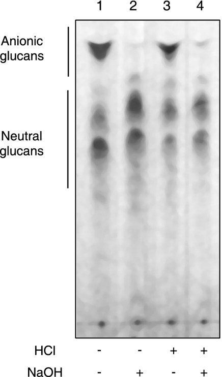
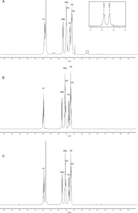
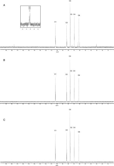
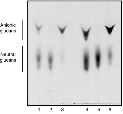
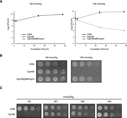
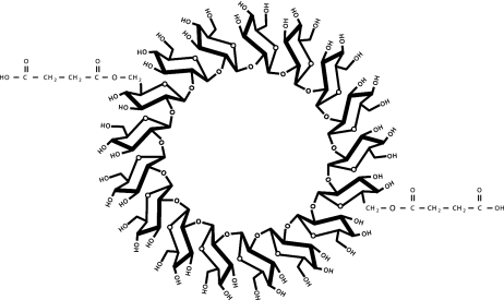
Brucella periplasmic cyclic beta-1,2-glucan plays an important role during bacterium-host interaction. Nuclear magnetic resonance spectrometry analysis, thin-layer chromatography, and DEAE-Sephadex chromatography were used to characterize Brucella abortus cyclic glucan. In the present study, we report that a fraction of B. abortus cyclic beta-1,2-glucan is substituted with succinyl residues, which confer anionic character on the cyclic beta-1,2-glucan. The oligosaccharide backbone is substituted at C-6 positions with an average of two succinyl residues per glucan molecule. This O-ester-linked succinyl residue is the only substituent of Brucella cyclic glucan. A B. abortus open reading frame (BAB1_1718) homologous to Rhodobacter sphaeroides glucan succinyltransferase (OpgC) was identified as the gene encoding the enzyme responsible for cyclic glucan modification. This gene was named cgm for cyclic glucan modifier and is highly conserved in Brucella melitensis and Brucella suis. Nucleotide sequencing revealed that B. abortus cgm consists of a 1,182-bp open reading frame coding for a predicted membrane protein of 393 amino acid residues (42.7 kDa) 39% identical to Rhodobacter sphaeroides succinyltransferase. cgm null mutants in B. abortus strains 2308 and S19 produced neutral glucans without succinyl residues, confirming the identity of this protein as the cyclic-glucan succinyltransferase enzyme. In this study, we demonstrate that succinyl substituents of cyclic beta-1,2-glucan of B. abortus are necessary for hypo-osmotic adaptation. On the other hand, intracellular multiplication and mouse spleen colonization are not affected in cgm mutants, indicating that cyclic-beta-1,2-glucan succinylation is not required for virulence and suggesting that no low-osmotic stress conditions must be overcome during infection.


Figures

FIG. 1.
FIG. 1.
Bio-Gel P4 chromatography of cyclic β-1,2-glucans accumulated in vivo by wild-type and mutant strains. Cells from 100-ml cultures were harvested and extracted with 70% ethanol. The extracts were subjected to chromatography on Bio-Gel P4 columns (78 by 1.8 cm). The glucans eluted with 0.1 M pyridine-acetate buffer (pH 5.5). Fractions of 1.5 ml were collected, and carbohydrates were expressed as μmol glucose equivalents (g wet weight)−1. (A) B. abortus 2308 cellular glucans. (B) B. abortus 2308 cellular glucans after mild alkali treatment (0.1 N NaOH for 30 min at 37°C). (C) B. abortus Cgm08 cellular glucans. (D) B. abortus Cgm(pBB4cgm) cellular glucans. V0, void volume; Vt, total volume; I, anionic glucan; II, neutral glucan.
FIG. 2.
FIG. 2.
DEAE-Sephadex chromatography of cellular glucans accumulated by wild-type and mutant strains. Glucans were recovered from Bio-Gel P4 columns (fractions 13 to 22) and subjected to chromatography on DEAE-Sephadex columns (0.25 by 3 cm). The glucans were eluted with 1.5 ml water (fractions 1 to 3), 1.5 ml 0.05 M NaCl (fractions 4 to 6), 1.5 ml 0.1 M NaCl (fractions 7 to 9), 1.5 ml 0.25 M NaCl (fractions 10 to 12), and 0.5 M NaCl (fractions 13 to 15). Fractions of 0.5 ml were collected, and carbohydrates were measured by the anthrone-sulfuric acid method (20). (A) B. abortus 2308 cellular glucans. (B) B. abortus 2308 cellular glucans after mild alkali treatment (0.1 N NaOH for 30 min at 37°C). (C) B. abortus Cgm08 cellular glucans. (D) B. abortus Cgm08(pBB4cgm) cellular glucans.
FIG. 3.
FIG. 3.
Thin-layer chromatographic analysis of ethanol extracts of B. abortus 2308. Ethanolic extracts were either directly applied to thin-layer chromatography plates (lane 1) or first subjected to mild alkali treatment (lane 2), acidic treatment (lane 3), or both treatments (lane 4). Alkali treatment resulted in selective removal of O-ester linkages.
FIG. 4.
FIG. 4.
1H-NMR spectra of purified cyclic β-1,2-glucans from B. abortus. (A) B. abortus S19. (B) B. abortus S19 treated with alkali. (C) B. abortus Cgm19. The two signals between 2.4 and 2.6 ppm correspond to the methylene protons of the succinic acid substituents. In panel A, the resonance at 3.16 is derived from methanol. The resonance at 4.7 is H2O. Chemical shifts are expressed relative to external 1,4-dioxane.
FIG. 5.
FIG. 5.
13C-NMR spectra of purified cyclic β-1,2-glucans from B. abortus. (A) B. abortus S19. (B) B. abortus S19 treated with alkali. (C) B. abortus Cgm19. Chemical shifts are expressed relative to external 1,4-dioxane (δ 67.4 ppm).
FIG. 6.
FIG. 6.
TLC of cyclic β-1,2-glucans formed by B. abortus strains. Total cellular glucans of B. abortus strains were extracted and subjected to TLC as described in Materials and Methods. B. abortus S19 (lanes 1 to 3): lane 1, wild type; lane 2, Cgm19; lane 3, Cgm19(pBB4cgm). B. abortus 2308 (lanes 4 to 6): lane 4, wild-type; lane 5, Cgm08; lane 6, Cgm08(pBB4cgm).
FIG. 7.
FIG. 7.
Effects of osmolarity on growth of different strains of B. abortus. (A) Studies in liquid media. B. abortus 2308 wild type, Cgm08, and the complemented strain Cgm08(pBB4cgm) were grown in LB-NaCl or LB as described in Materials and Methods. The number of viable bacteria at each time point was determined by making serial dilutions in PBS and plating aliquots on brucella broth plates. (B and C) Studies on solid media. For studies on solid media, 10 μl of the dilutions were spotted on LB agar or LB agar with the addition of 170 mM NaCl (B) or on LB agar or LB agar with one of the following additions: 170 mM KCl, 110 mM K2SO4, or 340 mM sucrose (C). The plates were incubated at 37°C for 5 days before being read.
FIG. 8.
FIG. 8.
Schematic representation of the structure of the succinyl-substituted cyclic β-1,2-glucan (degree of polymerization, 17) synthesized by B. abortus.

References

    1. Altabe, S. G., P. Talaga, J. M. Wieruszeski, G. Lippens, R. Ugalde, and J. P. Bohin. 1998. Periplasmic glucans of Azospirillum brasilense, p. 390. In C. Elmerich, A. Kondorosi, and W. E. Newton (ed.), Biological nitrogen fixation for the 21st century. Kluwer Academic Publishers, Dordrecht, The Netherlands.
    1. Arellano-Reynoso, B., N. Lapaque, S. Salcedo, G. Briones, A. E. Ciocchini, R. Ugalde, E. Moreno, I. Moriyon, and J. P. Gorvel. 2005. Cyclic beta-1,2-glucan is a Brucella virulence factor required for intracellular survival. Nat. Immunol. 6:618-625. - PubMed
    1. Batley, M., J. W. Redmond, and A. J. Wicken. 1987. Nuclear magnetic resonance spectra of lipoteichoic acid. Biochim. Biophys. Acta 901:127-137. - PubMed
    1. Bohin, J. P. 2000. Osmoregulated periplasmic glucans in Proteobacteria. FEMS Microbiol. Lett. 186:11-19. - PubMed
    1. Bohin, J. P., and E. P. Kennedy. 1984. Regulation of the synthesis of membrane-derived oligosaccharides in Escherichia coli. Assay of phosphoglycerol transferase I in vivo. J. Biol. Chem. 259:8388-8393. - PubMed

Publication types

MeSH terms

Associated data

LinkOut - more resources