. 2006 Jul;188(14):5273-85.
doi: 10.1128/JB.00300-06.

Modulation of the ComA-dependent quorum response in Bacillus subtilis by multiple Rap proteins and Phr peptides

Affiliations

Modulation of the ComA-dependent quorum response in Bacillus subtilis by multiple Rap proteins and Phr peptides

Jennifer M Auchtung et al. J Bacteriol. 2006 Jul.

Abstract

In Bacillus subtilis, extracellular peptide signaling regulates several biological processes. Secreted Phr signaling peptides are imported into the cell and act intracellularly to antagonize the activity of regulators known as Rap proteins. B. subtilis encodes several Rap proteins and Phr peptides, and the processes regulated by many of these Rap proteins and Phr peptides are unknown. We used DNA microarrays to characterize the roles that several rap-phr signaling modules play in regulating gene expression. We found that rapK-phrK regulates the expression of a number of genes activated by the response regulator ComA. ComA activates expression of genes involved in competence development and the production of several secreted products. Two Phr peptides, PhrC and PhrF, were previously known to stimulate the activity of ComA. We assayed the roles that PhrC, PhrF, and PhrK play in regulating gene expression and found that these three peptides stimulate ComA-dependent gene expression to different levels and are all required for full expression of genes activated by ComA. The involvement of multiple Rap proteins and Phr peptides allows multiple physiological cues to be integrated into a regulatory network that modulates the timing and magnitude of the ComA response.


Figures

FIG. 1.
FIG. 1.
Overexpression of rapF or rapK and deletion of phrC, phrF, or phrK inhibit expression of genes activated by ComA. We used DNA microarrays to examine changes in mRNA levels in response to overexpression of rapF, rapH, rapJ, or rapK or deletion of phrC, phrF, or phrK. A. Genes whose expression changed significantly in response to overexpression of rapF (RapF++), rapJ (RapJ++), or rapK (RapK++), to cooverexpression of rapH and phrH (RapH++), or to deletion of phrC (PhrC−), phrF (PhrF−), or phrK (PhrK−) were identified as described in Materials and Methods and are represented by a box shaded to represent the magnitude of the mean fold change in gene expression. A threefold or greater decrease in gene expression is shaded bright blue, and a threefold or greater increase in gene expression is shaded bright yellow. Those genes whose expression did not change significantly are shaded black. Additional microarray results, including the gene names and numerical values of the fold changes in gene expression, are in Table S1 in the supplemental material. The boxes to the left of the visualization indicate those genes whose expression were previously shown to be regulated by the response regulators ComA (5, 9, 53, 54) (gray box), Spo0A (14, 46) (black boxes), and DegU (40, 54) (white boxes). B to F. Percentage of operons whose expression changed significantly in response to overexpression of rapF or rapK or to deletion of phrC, phrF, or phrK and are known to be regulated by the response regulators ComA (green segments), Spo0A (striped segments), DegU (red segments), or other regulators (gray segments). B. Gene expression changes in cells overexpressing rapF (RapF++). C. Gene expression changes in cells overexpressing rapK (RapK++). D. Gene expression changes in ΔphrC cells (PhrC−). E. Gene expression changes in ΔphrF cells (PhrF−). F. Gene expression changes in ΔphrK cells (PhrK−).
FIG. 2.
FIG. 2.
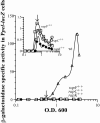
Overexpression of rapC, rapF, or rapK inhibits expression of the ComA-activated gene pel. Cells containing Ppel-lacZ and Pspank(hy)-rapC (JMA78), Pspank(hy)-rapF (JMA76), Pspank(hy)-rapK (JMA77), or Pspank(hy) (JMA79) were grown in defined minimal medium. IPTG was added to cells at an OD600 of ∼0.4 to 0.6. Samples were collected from cells prior to IPTG addition, at the time of IPTG addition, and 30, 60, 90 120, 150, and 180 min. after IPTG addition. β-Galactosidase activity was assayed as described in Materials and Methods and is plotted relative to the OD600 values of the samples. The arrow indicates the time of IPTG addition. Ppel-lacZ expression in Pspank(hy) (▵) (wild type [wt]), Pspank(hy)-rapC (○) (rapC++), Pspank(hy)-rapF (□) (rapF++), and Pspank(hy)-rapK (♦) (rapK++) cells is shown. The inset shows data replotted with the y axis from 0 to 1.5 units of β-galactosidase specific activity.
FIG. 3.
FIG. 3.
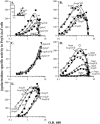
Effects of rap and phr deletions on expression of srfA. PsrfA-lacZ-containing cells were grown in defined minimal medium, and samples were removed throughout growth for determination of β-galactosidase specific activity. β-Galactosidase specific activity was determined as described in Materials and Methods and is plotted relative to the OD600 values of the samples. A. Wild-type (wt) (JMS682) (▪), ΔphrK (CAL8) (⋄), ΔphrC (JMA165) (○), ΔphrC ΔphrKphrCK) (CAL9) (•), and ΔphrF (JMA 166) (▵) cells. The inset shows data from the early time points (OD600 of <1) in wild-type, ΔphrC, and ΔphrF cells replotted with a y axis from 0 to 150 units of β-galactosidase specific activity. B. Wild-type (JMS682) (▪), ΔrapC (JMA47) (⋄), Δ(rapF phrF) (ΔrapF) (JMA117) (▴), ΔrapK (JMA48) (○), and ΔrapC Δ(rapF phrF) ΔrapKrapCFK) (JMA142) (•) cells. C. ΔphrF (▵) (JMA166), ΔphrC ΔphrFphrCF) (JMA169) (♦), ΔphrF ΔphrKphrFK) (CAL10) (▾), and ΔphrC ΔphrF ΔphrKphrCFK) (CAL11) (□) cells. D. Wild-type (JMA682) (▪), Δopp (JMA52) (⋄), Δopp Δ(rapF phrF) (Δopp ΔrapF) (JMA122) (•), Δopp ΔrapC Δ(rapF phrF) (Δopp ΔrapCF) (JMA134) (♦), Δopp ΔrapC ΔrapKopp ΔrapCK) (JMA58) (▴), Δopp Δ(rapF phrF) ΔrapKopp ΔrapFK) (JMA138) (▵), and Δopp ΔrapC Δ(rapF phrF) ΔrapKopp ΔrapCFK) (JMA144) (□) cells. E. Wild-type (JMS682) (▪), ΔsigH (JMA51) (•), ΔsigH ΔrapC (JMA53) (⋄), ΔsigH Δ(rapF phrF) ΔrapKsigH ΔrapFK) (JMA139) (▿), and ΔsigH ΔrapC Δ(rapFphrF) ΔrapKsigH ΔrapCFK) (JMA149) (○) cells.
FIG. 4.
FIG. 4.
Integration of multiple signals for complex regulation of ComA-dependent gene expression. This diagram illustrates several of the physiological cues that are known to influence ComA-dependent gene expression. Further details are provided in the text.

References

    1. Ansaldi, M., D. Marolt, T. Stebe, I. Mandic-Mulec, and D. Dubnau. 2002. Specific activation of the Bacillus quorum-sensing systems by isoprenylated pheromone variants. Mol. Microbiol. 44:1561-1573. - PubMed
    1. Auchtung, J. M., C. A. Lee, R. E. Monson, A. P. Lehman, and A. D. Grossman. 2005. Regulation of a Bacillus subtilis mobile genetic element by intercellular signaling and the global DNA damage response. Proc. Natl. Acad. Sci. USA 102:12554-12559. - PMC - PubMed
    1. Bacon Schneider, K., T. M. Palmer, and A. D. Grossman. 2002. Characterization of comQ and comX, two genes required for production of ComX pheromone in Bacillus subtilis. J. Bacteriol. 184:410-419. - PMC - PubMed
    1. Benson, A. K., and W. G. Haldenwang. 1993. Regulation of sigma-B levels and activity in Bacillus subtilis. J. Bacteriol. 175:2347-2356. - PMC - PubMed
    1. Berka, R. M., J. Hahn, M. Albano, I. Draskovic, M. Persuh, X. Cui, A. Sloma, W. Widner, and D. Dubnau. 2002. Microarray analysis of the Bacillus subtilis K-state: genome-wide expression changes dependent on ComK. Mol. Microbiol. 43:1331-1345. - PubMed

Publication types

Associated data