. 2006 Jul;169(1):47-60.
doi: 10.2353/ajpath.2006.050975.

Obligatory role for interleukin-13 in obstructive lesion development in airway allografts

Affiliations

Obligatory role for interleukin-13 in obstructive lesion development in airway allografts

Vibha N Lama et al. Am J Pathol. 2006 Jul.

Abstract

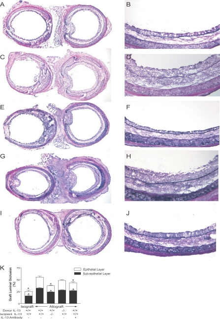
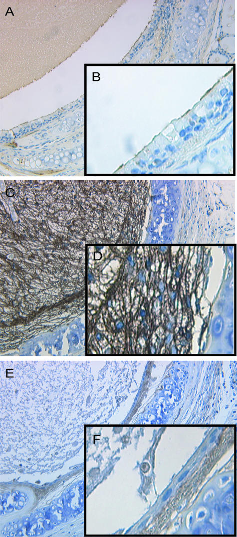
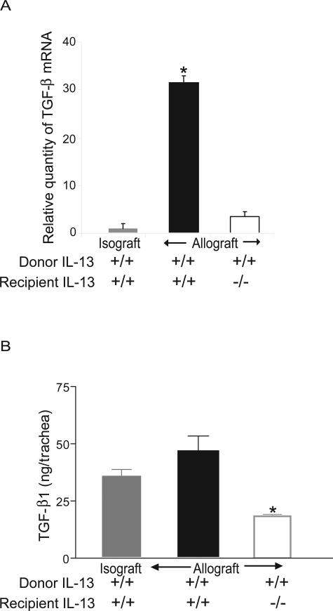
The pathogenesis of bronchiolitis obliterans (BO), a common and devastating obliterative disorder of small airways following lung transplantation, remains poorly understood. Lesions are characterized in their early stages by lymphocyte influx that evolves into dense fibrotic infiltrates. Airway specimens taken from patients with histological BO revealed infiltrating myofibroblasts, which strongly expressed the signaling chain of the high affinity interleukin-13 (IL-13) receptor IL-13Ralpha1. Because IL-13 has proinflammatory and profibrotic actions, a contributory role for IL-13 in BO development was examined using murine models of orthotopic and heterotopic tracheal transplantation. Compared with airway isografts, allografts exhibited a significant increase in relative IL-13 mRNA and protein levels. Allogeneic tracheas transplanted into IL-13-deficient mice were protected from BO in both transplant models. Flow cytometric analysis of orthotopic transplant tissue digests revealed markedly fewer infiltrating mononuclear phagocytes and CD3(+) T lymphocytes in IL-13-deficient recipients. Furthermore, protection from luminal obliteration, collagen deposition, and myofibroblast infiltration was observed in heterotopic airways transplanted into the IL-13(-/-) recipients. Transforming growth factor-beta1 expression was significantly decreased in tracheal allografts into IL-13(-/-) recipients, compared to wild-type counterparts. These human and murine data implicate IL-13 as a critical effector cytokine driving cellular recruitment and subsequent fibrosis in clinical and ex-perimental BO.


Figures

Figure 1
Figure 1
Immunohistochemistry staining for α-SMA and IL-13Rα1 in TBBx specimens demonstrating evidence of BO. A: α-SMA-positive (stained brown) mesenchymal cells (myofibroblasts) are demonstrated in the submucosa of bronchi in a patient with BOS. B: TBBx specimen from a patient without BOS showing normal bronchial epithelium with underlying smooth muscle bundle and absence of myofibroblast infiltration in the lamina propria and the lumen. C: α-SMA staining (brown) of a TBBx specimen demonstrates bronchi cut tangentially as evidenced by smooth muscle bundles (arrows). The lumen is filled with mesenchymal cells that stain positively for α-SMA, demonstrating the presence of myofibroblasts. D–F: Contiguous sections from C (marked by the rectangle) are shown at higher magnification stained with α-SMA (D), IL-13Rα1 (stained red) (E), and the negative control for IL-13Rα1 (F). Original magnifications: ×100 (A and C); ×200 (B); ×400 (D–F). These findings are representative of all 15 patients examined.
Figure 2
Figure 2
IL-13 up-regulation in the pathogenesis of murine BOS. A–D: Time-course analysis of up-regulation of IL-13 in BO. Real-time quantitative PCR of IL-13 in orthotopic (A) and heterotopic (C) tracheal transplants at 3, 7, 14, 21, and 28 days after transplantation. IL-13 mRNA was markedly elevated in allografts transplanted into BALB/c recipients compared to isografts at day 7 in both orthotopic (P = 0.0001) and heterotopic (P < 0.0001) models and continued to be elevated at day 14 in the heterotopic model (P < 0.0001). Representative band for IL-13 mRNA and β-actin, generated by RT-PCR, comparing isografts to allografts at day 7 are demonstrated for both the orthotopic (B) and the heterotopic (D) transplant models. E and F: Protein expression by ELISA in orthotopic (E) and heterotopic (F) tracheal transplant models demonstrates increased IL-13 protein in allografts compared to isografts. G and H: IL-13 mRNA expression in tracheal allografts transplanted into IL-13−/− animals, by real-time quantitative PCR, demonstrated no up-regulation of IL-13 at day 7 in both orthotopic (G) and heterotopic (H) models. Data shown represent mean ± SE for each group. *P < 0.05 compared to isografts. n = 4 tracheas per group for mRNA analysis. n = 4 tracheal homogenates of 2 tracheas per group for ELISA.
Figure 3
Figure 3
Analysis of graft luminal narrowing 1 week after transplantation in the orthotopic tracheal transplant model. Representative histology and morphometric analysis for each of the indicated conditions is shown. A and B: BALB/c into BALB/c isografts. C and D: C57Bl/6 allografts into BALB/c. E and F: C57Bl/6 allografts into IL-13−/− recipients. G and H: IL-13−/− allografts into C57Bl/6 recipients. I and J: C57Bl/6 allografts into BALB/c treated with IL-13 antibody. In A, C, E, G, and I the native trachea is shown on the left, and the orthotopic transplant is shown on the right. Data represent the analysis of at least four transplants per group. K: Quantitative morphometric analysis of tissue sections. Allografts treated with IL-13 antibody and allografts placed in IL-13−/− recipients were significantly protected compared to BALB/c recipients. *P < 0.05 compared to allografts. All allograft conditions were statistically different from the isograft.
Figure 4
Figure 4
FACS analysis and immunohistochemical staining for macrophages and T cells in orthotopic tracheal transplant model. A: FACS analysis of F4/80+ cells (macrophages) identified in cell isolations from allografts transplanted into BALB/c or IL-13−/− recipients. Markedly fewer infiltrating mononuclear phagocytes were identified in IL-13−/− recipients (P < 0.05). B: Immunohistochemical staining for F4/80+ in allografts transplanted into BALB/c mice. C: Staining for F4/80+ in an allograft transplanted into an IL-13−/− recipients. D: FACS analysis for CD3+ cells in cell isolations from allografts into BALB/c or IL-13−/− mice. Significantly decreased infiltration by CD3-positive lymphocytes was demonstrated in IL-13−/− allograft recipients as compared to wild-type recipients (P < 0.05). E: Staining for CD3 in allografts transplanted into BALB/c mice. F: Staining for CD3 in allografts transplanted into IL-13−/− recipients. All data represent analysis at 1 week after transplant. IL-13−/− recipients demonstrate significantly reduced recruitment of both macrophages and T cells (n = 4).
Figure 5
Figure 5
Histopathology (hematoxylin and eosin) of luminal fibrosis 28 days after transplantation in the heterotopic tracheal transplant model. A and B: Representative photomicrographs of heterotopic trachea allografts (C57Bl/6 allografts into BALB/c-recipient mice) demonstrate epithelial injury, complete airway obliteration, and matrix deposition. C and D: Allografts into IL-13−/−-recipient mice show intact epithelium and no intraluminal cellular infiltration or airway obliteration. The deposit in the center represents acellular debris as this is not an air-permissive model. E and F: IL-13−/− tracheas used as donors into C57Bl/6 mice do not demonstrate any protection from airway obliteration. G: Quantitative analysis of histopathological sections of tracheal allografts transplanted into IL-13+/+ and IL-13−/− recipients. Histology shown is representative of n = 4–6 per group. Original magnifications: ×200 (A, C, and E); ×400 (B, D, and F). *P < 0.05.
Figure 6
Figure 6
Trichrome staining and quantification of collagen deposition in heterotopic tracheal allografts transplanted into IL-13−/− and wild-type mice. A and D: Representative photomicrographs of Masson’s trichrome staining (blue color) for collagen demonstrates early intraluminal collagen deposition in mouse allografts. B and E: No significant intraluminal collagen was seen in allografts transplanted into IL-13−/−-recipient mice. C and F: IL-13−/− mice as donors were indistinguishable from control allografts. Histology shown is representative of n = 4–6 per group. G: Quantification of collagen deposition by the Sircol assay at day 28 demonstrates significantly increased level of collagen in allografts as compared to isografts (P < 0.01). However, tracheal allografts placed into IL-13−/− mice were significantly protected from collagen deposition (P < 0.001 for allografts into IL-13−/− mice compared with allografts into BALB/c mice). No significant difference in collagen levels was seen between isografts and IL-13−/− allografts (P > 0.05). Values represent the mean ± SE of six tracheal transplants per group. *P < 0.05 versus isograft.
Figure 7
Figure 7
Immunohistochemical staining for α-SMA in tracheal transplants. A and B: α-SMA staining (brown) in a representative isograft demonstrates intact epithelium and only smooth muscle cell staining. The intraluminal brown staining represents acellular debris. C and D: Allografts demonstrate intraluminal mesenchymal cells, which stain intensely for α-SMA obliterating the lumen, thus demonstrating myofibroblast accumulation. E and F: Allografts into IL-13−/− mice demonstrate myofibroblast infiltration restricted to lamina propria with intact epithelium. Acellular amorphous debris is seen in the lumen. Histology shown is representative of n = 4 per group. Original magnifications: ×200 (A, C, and E); ×400 (B, D, and F).
Figure 8
Figure 8
TGF-β1 mRNA expression and bioactivity in heterotopic tracheal allografts. A: TGF-β1 mRNA expression by real-time quantitative PCR in tracheal transplants at day 28 demonstrates a 32-fold increase in TGF-β1 in control allografts as compared to isografts (P < 0.001). However, no significant increase in TGF-β1 mRNA expression was seen in allograft tracheas transplanted into IL-13−/− mice as compared to isografts. B: Tracheal homogenates from isografts and allografts transplanted into BALB/c or IL-13−/− recipients were analyzed for TGF-β1 bioactivity by PAIL assay at day 28. TGF-β1 bioactivity was significantly lower in allografts transplanted into IL-13+/+ compared to IL-13−/− recipients (P < 0.001). Data represent the mean ± SE for four tracheas per group measured in duplicate. *P < 0.05.

Similar articles

Cited by

References

    1. Boehler A, Kesten S, Weder W, Speich R. Bronchiolitis obliterans after lung transplantation: a review. Chest. 1998;114:1411–1426. - PubMed
    1. Estenne M, Maurer JR, Boehler A, Egan JJ, Frost A, Hertz M, Mallory GB, Snell GI, Yousem S. Bronchiolitis obliterans syndrome 2001: an update of the diagnostic criteria. J Heart Lung Transplant. 2002;21:297–310. - PubMed
    1. Sharples LD, McNeil K, Stewart S, Wallwork J. Risk factors for bronchiolitis obliterans: a systematic review of recent publications. J Heart Lung Transplant. 2002;21:271–281. - PubMed
    1. Hertz MI, Jessurun J, King MB, Savik SK, Murray JJ. Reproduction of the obliterative bronchiolitis lesion after heterotopic transplantation of mouse airways. Am J Pathol. 1993;142:1945–1951. - PMC - PubMed
    1. Boehler A, Chamberlain D, Kesten S, Slutsky AS, Liu M, Keshavjee S. Lymphocytic airway infiltration as a precursor to fibrous obliteration in a rat model of bronchiolitis obliterans. Transplantation. 1997;64:311–317. - PubMed

Publication types

MeSH terms