

Chronically increased transforming growth factor-beta1 strongly inhibits hippocampal neurogenesis in aged mice
- PMID: 16816369
- PMCID: PMC1698757
- DOI: 10.2353/ajpath.2006.051272
Chronically increased transforming growth factor-beta1 strongly inhibits hippocampal neurogenesis in aged mice
Abstract
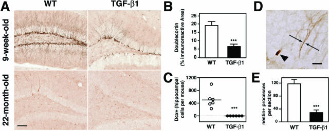
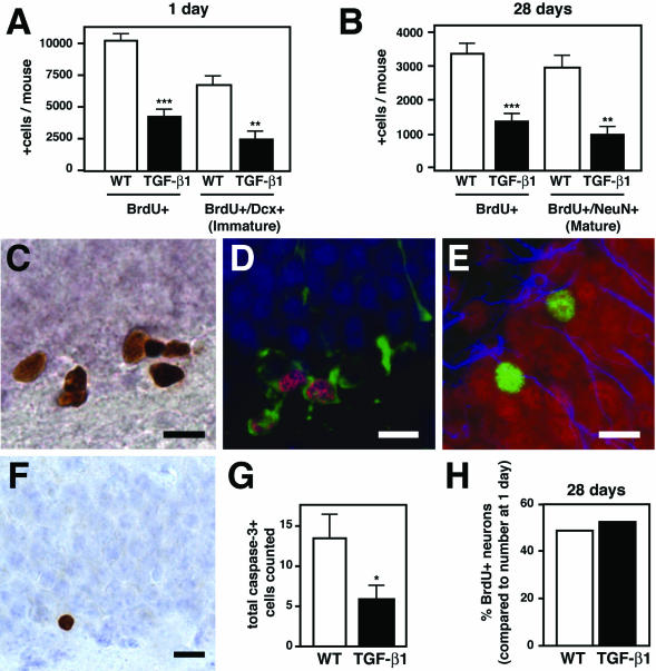
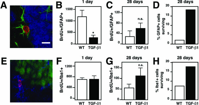
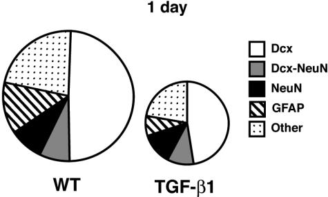
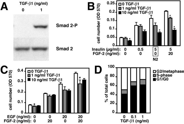
There is increasing evidence that hippocampal learning correlates strongly with neurogenesis in the adult brain. Increases in neurogenesis after brain injury also correlate with improved outcomes. With aging the capacity to generate new neurons decreases dramatically, both under normal conditions and after injury. How this decrease occurs is not fully understood, but we hypothesized that transforming growth factor (TGF)-beta1, a cell cycle regulator that rapidly increases after injury and with age, might play a role. We found that chronic overproduction of TGF-beta1 from astrocytes almost completely blocked the generation of new neurons in aged transgenic mice. Even young adult TGF-beta1 mice had 60% fewer immature, doublecortin-positive, hippocampal neurons than wild-type littermate controls. Bromodeoxyuridine labeling of dividing cells in 2-month-old TGF-beta1 mice confirmed this decrease in neuro-genesis and revealed a similar decrease in astrogenesis. Treatment of early neural progenitor cells with TGF-beta1 inhibited their proliferation. This strongly suggests that TGF-beta1 directly affects these cells before their differentiation into neurons and astrocytes. Together, these data show that TGF-beta1 is a potent inhibitor of hippocampal neural progenitor cell proliferation in adult mice and suggest that it plays a key role in limiting injury and age-related neurogenesis.
Figures
Similar articles
-
TGF-beta signalling in the adult neurogenic niche promotes stem cell quiescence as well as generation of new neurons.J Cell Mol Med. 2014 Jul;18(7):1444-59. doi: 10.1111/jcmm.12298. Epub 2014 Apr 30. J Cell Mol Med. 2014. PMID: 24779367 Free PMC article.
-
Transforming growth factor-beta1 is a negative modulator of adult neurogenesis.J Neuropathol Exp Neurol. 2006 Apr;65(4):358-70. doi: 10.1097/01.jnen.0000218444.53405.f0. J Neuropathol Exp Neurol. 2006. PMID: 16691117
-
Neuro-glia interaction effects on GFAP gene: a novel role for transforming growth factor-beta1.Eur J Neurosci. 2002 Dec;16(11):2059-69. doi: 10.1046/j.1460-9568.2002.02283.x. Eur J Neurosci. 2002. PMID: 12473073
-
Research update: neurogenesis in adult brain and neuropsychiatric disorders.Mt Sinai J Med. 2006 Nov;73(7):931-40. Mt Sinai J Med. 2006. PMID: 17195878 Review.
-
Hippocampal neurogenesis during normal and pathological aging.Psychoneuroendocrinology. 2007 Aug;32 Suppl 1:S26-30. doi: 10.1016/j.psyneuen.2007.04.014. Epub 2007 Jul 12. Psychoneuroendocrinology. 2007. PMID: 17629417 Review.
Cited by
-
Neuronal Rac1 is required for learning-evoked neurogenesis.J Neurosci. 2013 Jul 24;33(30):12229-41. doi: 10.1523/JNEUROSCI.2939-12.2013. J Neurosci. 2013. PMID: 23884931 Free PMC article.
-
Surveillance, phagocytosis, and inflammation: how never-resting microglia influence adult hippocampal neurogenesis.Neural Plast. 2014;2014:610343. doi: 10.1155/2014/610343. Epub 2014 Mar 19. Neural Plast. 2014. PMID: 24772353 Free PMC article. Review.
-
Control of Cell Survival in Adult Mammalian Neurogenesis.Cold Spring Harb Perspect Biol. 2015 Oct 28;7(12):a018895. doi: 10.1101/cshperspect.a018895. Cold Spring Harb Perspect Biol. 2015. PMID: 26511628 Free PMC article. Review.
-
m6A/YTHDF2-mediated mRNA decay targets TGF-β signaling to suppress the quiescence acquisition of early postnatal mouse hippocampal NSCs.Cell Stem Cell. 2025 Jan 2;32(1):144-156.e8. doi: 10.1016/j.stem.2024.10.002. Epub 2024 Oct 29. Cell Stem Cell. 2025. PMID: 39476834
-
Transgenic models for cytokine-induced neurological disease.Biochim Biophys Acta. 2010 Oct;1802(10):903-17. doi: 10.1016/j.bbadis.2009.10.004. Epub 2009 Oct 14. Biochim Biophys Acta. 2010. PMID: 19835956 Free PMC article. Review.
References
-
- Palmer TD, Willhoite AR, Gage FH. Vascular niche for adult hippocampal neurogenesis. J Comp Neurol. 2000;425:479–494. - PubMed
-
- Shors TJ, Miesegaes G, Beylin A, Zhao M, Rydel T, Gould E. Neurogenesis in the adult is involved in the formation of trace memories. Nature. 2001;410:372–376. - PubMed
-
- Rola R, Raber J, Rizk A, Otsuka S, VandenBerg SR, Morhardt DR, Fike JR. Radiation-induced impairment of hippocampal neurogenesis is associated with cognitive deficits in young mice. Exp Neurol. 2004;188:316–330. - PubMed
Publication types
MeSH terms
Substances
Grants and funding
LinkOut - more resources
Full Text Sources
Medical
Molecular Biology Databases
