Comparative Study
. 2006 Jul;169(1):189-99.
doi: 10.2353/ajpath.2006.051225.

Role of neuronal interferon-gamma in the development of myelopathy in rats infected with human T-cell leukemia virus type 1

Affiliations
Comparative Study

Role of neuronal interferon-gamma in the development of myelopathy in rats infected with human T-cell leukemia virus type 1

Yukiko Miyatake et al. Am J Pathol. 2006 Jul.

Abstract

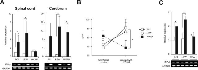
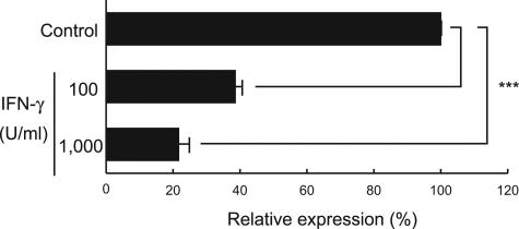
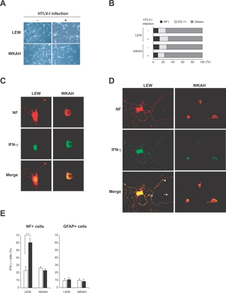
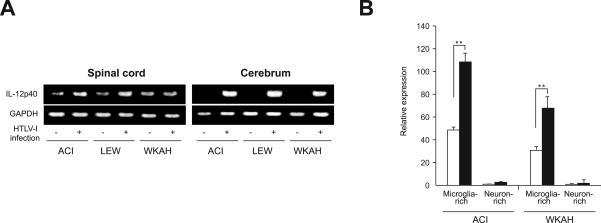
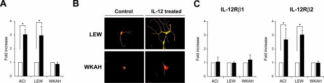
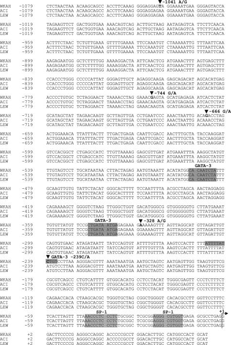
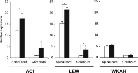
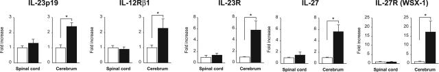
Human T-cell leukemia virus type 1 (HTLV-1) is the causative agent of not only adult T-cell leukemia but also HTLV-1-associated myelopathy/tropical spastic paraparesis (HAM/TSP). Among the rat strains infected with HTLV-1, chronic progressive myelopathy, named HAM rat disease, occurs exclusively in WKAH rats. In the present study, we found that HTLV-1 infection induces interferon (IFN)-gamma production in the spinal cords of HAM-resistant strains but not in those of WKAH rats. Neurons were the major cells that produced IFN-gamma in HTLV-1-infected, HAM-resistant strains. Administration of IFN-gamma suppressed expression of pX, the gene critically involved in the onset of HAM rat disease, in an HTLV-1-immortalized rat T-cell line, indicating that IFN-gamma protects against the development of HAM rat disease. The inability of WKAH spinal cord neurons to produce IFN-gamma after infection appeared to stem from defects in signaling through the interleukin (IL)-12 receptor. Specifically, WKAH-derived spinal cord cells were unable to up-regulate the IL-12 receptor beta2 gene in response to IL-12 stimulation. We suggest that the failure of spinal cord neurons to produce IFN-gamma through the IL-12 pathway is involved in the development of HAM rat disease.


Figures

Figure 1
Figure 1
A: The amount of IFN-γ mRNA in the spinal cord and cerebrum was quantified by real-time RT-PCR. Samples were obtained from rats 7 months after HTLV-1 infection (black columns) and from age-matched uninfected controls (white columns). Results of experiments done in triplicate were evaluated as relative expression levels to the GAPDH gene. Data are represented as mean ± SD values of experiments done independently three times. Representative photos of gel electrophoresis of RT-PCR products are shown beneath the graph. B: The amount of IFN-γ protein in the spinal cord was quantified using the ELISA kit. Samples were obtained from rats 7 months after HTLV-1 infection and from age-matched uninfected controls. Data are represented as mean ± SD values of experiments done independently three times. C: The amount of IRF-1 mRNA in the spinal cord was quantified by real-time RT-PCR. Samples were obtained from rats 7 months after HTLV-1 infection (black columns) and from age-matched uninfected controls (white columns). Results of experiments done in triplicate were evaluated as relative expression levels to the GAPDH gene. Data are represented as mean ± SD values of experiments done independently three times. Representative photos of gel electrophoresis of RT-PCR products are shown beneath the graph. In each group of all experiments, at least three rats were used. *P < 0.05.
Figure 2
Figure 2
The HTLV-1-immortalized rat T-cell line, LEW-S1, was incubated with 100 or 1000 U/ml of recombinant rat IFN-γ for 3 hours, and then expression of the pX gene was quantified using the real-time RT-PCR method. Results of experiments done in triplicate were evaluated as relative expression levels to the structural gag gene. Data are represented as mean ± SD values of experiments done independently three times. ***P < 0.0001.
Figure 3
Figure 3
Spinal cord cells were isolated from HTLV-1-infected rats and from age-matched uninfected rats. Infected rats were sacrificed 7 months after inoculation with HTLV-1. A: Phase-contrast photos of cultured cells. B: Cells cultured on poly-l-lysine/laminin-coated glasses for 5 days were fixed and then used for double-immunofluorescent staining for NF (a marker for neurons) and ED-1 (a marker for microglia). The proportion of each cell population in the culture was calculated. C and D: Spinal cord cells derived from uninfected (C) and HTLV-1-infected rats (D) were stained for NF (red) and IFN-γ (green). Arrowheads indicate the expression of IFN-γ in synapses. E: Ratios of IFN-γ+ cells in neurons (NF+ cells) and astrocytes (GFAP+ cells) in the spinal cord. Cells were isolated from the spinal cord of rats 7 months after HTLV-1 infection (black columns) and from age-matched uninfected controls (white columns). After double staining for IFN-γ and NF, or IFN-γ and GFAP, 100 cells were counted in three independent high-power views, respectively, under a fluorescence microscope. In each group, at least three rats were used. Results are represented as mean ± SD values of experiments done independently three times. **P < 0.001. Original magnifications: ×200 (A); ×620 (C, D).
Figure 4
Figure 4
A: Expression of the IL-12p40 gene in the spinal cord and cerebrum. Samples were obtained from rats 7 months after HTLV-1 infection and from age-matched uninfected controls. Experiments were repeated independently at least three times. Representative photos of gel electrophoresis of RT-PCR products are shown. B: The amount of IL-12p40 mRNA in microglia- or neuron-rich populations prepared from the spinal cord was quantified by real-time RT-PCR. Samples were obtained from rats 7 months after HTLV-1 infection (black columns) and from age-matched uninfected controls (white columns). Data (relative expression levels to the GAPDH gene) are represented as mean ± SE values obtained from experiments performed in triplicate and repeated three times. In each group, at least three rats were used. **P < 0.001.
Figure 5
Figure 5
A: The amount of IFN-γ mRNA in the cells isolated from the spinal cord of uninfected rats was quantified by real-time RT-PCR. Samples were obtained from the cells after treatment with recombinant IL-12 (100 ng/ml) for 18 hours (black columns). Results of experiments done in triplicate and repeated three times were evaluated as mean ± SE values of the fold increase to the data without IL-12 treatment (white columns). *P < 0.05. B: Cells isolated from the spinal cord of uninfected rats were cultured on poly-l-lysine/laminin-coated glasses. After incubation with recombinant IL-12 (100 ng/ml) for 5 days, immunofluorescent double staining was performed using anti-IFN-γ (green) and anti-NF (red) antibodies. Representative merged images are shown. Experiments were performed independently three times. C: The amount of IL-12Rβ1 and IL-12Rβ2 mRNAs in the cells isolated from the spinal cord of uninfected rats was quantified by real-time RT-PCR. Samples were obtained from the cells after treatment with recombinant IL-12 (100 ng/ml) for 18 hours (black columns). Results of experiments done in triplicate and repeated three times were evaluated as mean ± SE values of the fold increase to the data without IL-12 treatment (white columns). *P < 0.05. For all experiments, at least three rats were used in each group. Original magnifications, ×620 (B).
Figure 6
Figure 6
The 5′-flanking region of the rat IL-12Rβ2 gene. The genomic DNA was extracted from the tail of HAM-susceptible (WKAH) and HAM-resistant (ACI, LEW) rats, and then the 5′-flanking region of the IL-12Rβ2 gene was amplified by nested PCR. The PCR products were purified and subjected to direct sequencing. Shaded sequences represent potential SP-1 or GATA-3 binding sites. +1 represents a tentatively assigned transcription start site deduced from the data available for the mouse IL-12Rβ2 gene.
Figure 7
Figure 7
The amount of IL-12Rβ2 mRNA in the spinal cord and cerebrum was quantified by real-time RT-PCR. Samples were obtained from rats 7 months after HTLV-1 infection (black columns) and from age-matched uninfected controls (white columns). Results of experiments performed in triplicate were evaluated as relative expression levels to the GAPDH gene. For each strain, the expression level of IL-12Rβ2 mRNA in the cerebrum of uninfected control rats was set as 1. Relative expression levels (mean ± SD values) were determined from the experiments done independently three times. In each group, at least three rats were used. *P < 0.05.
Figure 8
Figure 8
The amounts of IL-23p19, IL-12Rβ1, IL-23R, IL-27, and IL-27R (WSX-1) mRNAs in the spinal cord and cerebrum were quantified by real-time RT-PCR. Samples were obtained from WKAH rats 7 months after HTLV-1 infection (black columns) and from age-matched uninfected controls (white columns). In each group, at least three rats were used. Results of experiments done in triplicate were evaluated as relative expression levels to the GAPDH gene. Data from experiments done independently three times are represented as the fold increase (mean ± SD values) to the data without infection (white columns). *P < 0.05.

References

    1. Poiesz BJ, Ruscetti FW, Gazdar AF, Bunn PA, Minna JD, Gallo RC. Detection and isolation of type C retrovirus particles from fresh and cultured lymphocytes of a patient with cutaneous T-cell lymphoma. Proc Natl Acad Sci USA. 1980;77:7415–7419. - PMC - PubMed
    1. Yoshida M, Miyoshi I, Hinuma Y. Isolation and characterization of retrovirus from cell lines of human adult T-cell leukemia and its implication in the disease. Proc Natl Acad Sci USA. 1982;79:2031–2035. - PMC - PubMed
    1. Gessain A, Barin F, Vernant JC, Gout O, Maurs L, Calender A, de Thé G. Antibodies to human T-lymphotropic virus type-I in patients with tropical spastic paraparesis. Lancet. 1985;2:407–410. - PubMed
    1. Osame M, Usuku K, Izumo S, Ijichi N, Amitani H, Igata A, Matsumoto M, Tara M. HTLV-I associated myelopathy, a new clinical entity. Lancet. 1986;1:1031–1032. - PubMed
    1. Mochizuki M, Watanabe T, Yamaguchi K, Takatsuki K, Yoshimura K, Shirao M, Nakashima S, Mori S, Araki S, Miyata N. HTLV-I uveitis: a distinct clinical entity caused by HTLV-I. Jpn J Cancer Res. 1992;83:236–239. - PMC - PubMed

Publication types

MeSH terms

Associated data