. 2006 Jul;169(1):325-36.
doi: 10.2353/ajpath.2006.060206.

Involvement of endothelial CD44 during in vivo angiogenesis

Affiliations

Involvement of endothelial CD44 during in vivo angiogenesis

Gaoyuan Cao et al. Am J Pathol. 2006 Jul.

Erratum in

  • Am J Pathol. 2006 Nov;169(5):1899

Abstract

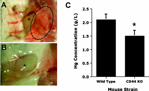
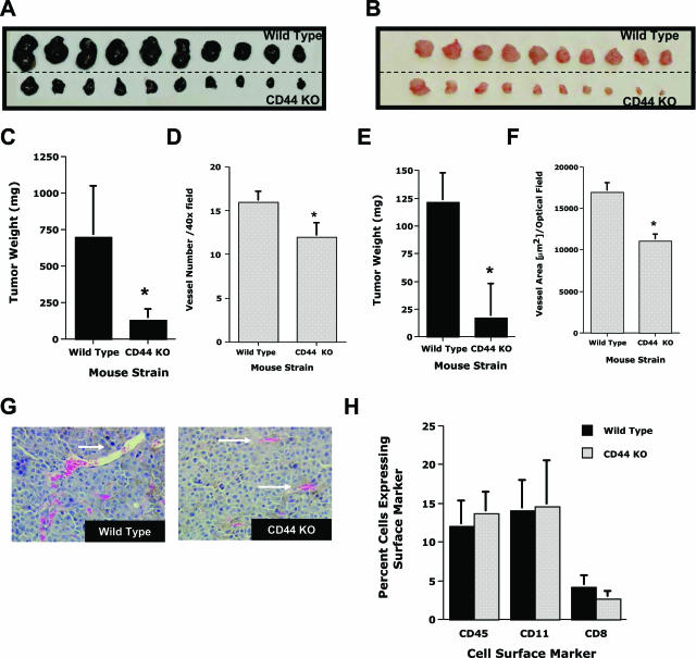
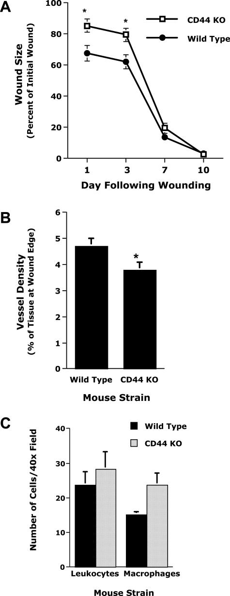
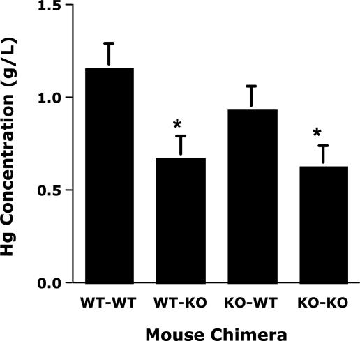
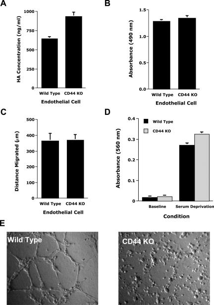
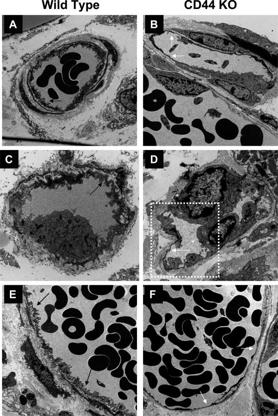
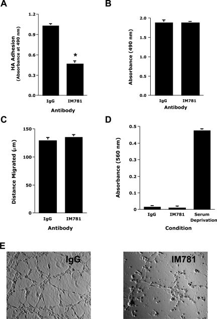
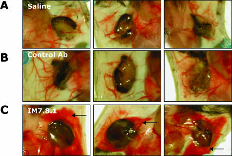
CD44, a cell-surface receptor for hyaluronan, has been implicated in endothelial cell functions, but its role in the formation of blood vessels in vivo has not been established. In CD44-null mice, vascularization of Matrigel implants and tumor and wound angiogenesis were inhibited. Leukocyte accumulation during tumor growth and wound healing in wild-type and CD44-null mice were comparable, and reconstitution of CD44-null mice with wild-type bone marrow did not restore the wild-type phenotype, suggesting that impairments in angiogenesis in CD44-deficient mice are due to the loss of endothelial CD44. Although the cell proliferation, survival, and wound-induced migration of CD44-null endothelial cells were intact, these cells were impaired in their in vitro ability to form tubes. Nascent vessels in Matrigel implants from CD44-null mice demonstrated irregular luminal surfaces characterized by retracted cells and thinned endothelia. Further, an anti-CD44 antibody that disrupted in vitro tube formation induced hemorrhage around Matrigel implants, suggesting that antagonism of endothelial CD44 undermined the integrity of the endothelium of nascent vessels. These data establish a role for CD44 during in vivo angiogenesis and suggest that CD44 may contribute to the organization and/or stability of developing endothelial tubular networks.


Figures

Figure 1
Figure 1
Neovascularization of Matrigel implants in wild-type and CD44-null mice. Shown are images of Matrigel implants (star) containing B16 melanoma cells as a source of angiogenic factors harvested after 5 days from wild-type (A) and CD44-null (CD44 KO) (B) animals. A “blush” of vessel proliferation (circle) was evident around the implants from the wild-type animal that was not present in the CD44-null mice. The vascularization of the gels, as assessed by hemoglobin concentration (C), was significantly reduced in CD44-null mice compared to wild-type mice. Data are presented as means ± SE (n = 20, *P = 0.02).
Figure 2
Figure 2
Tumor growth and angiogenesis in CD44-null mice. The growth of the B16 melanoma line (A and C) and the ID8-VEGF ovarian tumor line (B and E), as accessed by tumor weight, were significantly inhibited in the CD44-null (CD44 KO) mice (n = 10, *P < 0.01). The vessel densities of tumors of comparable sizes, as assessed by vessel number/40× field for the B16 tumor (D) and vessel area, μm2/optical field for the ID8-VEGF tumor (F), were significantly reduced in the CD44-null mice (n = 4–6, *P < 0.002). G: Hematoxylin and eosin sections of B16 melanomas from CD44-null mice revealed a paucity of the large vascular spaces (arrows) that were readily observable in the wild-type animals. H: The percentage of cells from B16 tumors expressing leukocyte surface markers (CD11b, CD45, or CD8) was determined by FACS analysis and found to be comparable in wild-type and CD44-null animals. Data are presented as means ± SE.
Figure 3
Figure 3
Wound healing and angiogenesis in CD44-null mice. A: The closure of 1-cm2 skin wounds, as measured by percentage of initial wound, was less at days 1 and 3 after wounding in CD44-null (CD44 KO) animals compared to wild-type mice, but not at later time points (n = 6, *P < 0.02). B: Vessel density at the edge of the wound (percentage of tissue at wound edge occupied by vessels) on day 3 was reduced (by 20%) in the CD44-null mice (B) (n = 8, *P < 0.03). C: Leukocyte and macrophage accumulations (number of cells/40× field) in the two mice strains were comparable (n = 4; *P > 0.1). Data are presented as means ± SE.
Figure 4
Figure 4
Neovascularization of Matrigel implants in bone marrow chimeric mice. Vascularization of Matrigel implants was studied in the following chimeric (donor-recipient) mice: wild-type into wild-type mice (WT-WT), wild-type into CD44-null mice (WT-KO), CD44-null into wild-type mice (KO-WT), and CD44-null into CD44-null mice (KO-KO). The angiogenic responses (assessed by hemoglobin concentration) in the WT-KO and KO-KO mice were very similar but were significantly reduced compared to the WT-WT and KO-WT responses, which were similar. Data are presented as means ± SE (n = 8, *P < 0.02).
Figure 5
Figure 5
In vitro function of murine ECs isolated from wild-type and CD44-null mice. A: Binding of HA to ECs isolated from wild-type and CD44-null (CD44 KO) mice was determined by enzyme-linked immunosorbent assay (n = 2). Before the addition of exogenous HA, the background levels of HA associated with cell surface of the wild-type and the CD44-null cells were 384 and 295 ng/ml, respectively. HA binding to CD44-null cells was preserved. B: The proliferation of wild-type and CD44-null ECs cultured for 24 hours in the presence of serum was assessed using a colorimetric assay and measurement of the reaction mixture at 490 nm. The proliferative responses of the two cell types were comparable (n = 4). C: Liner defects were made in confluent cell monolayers, and closure of the wounds after 24 hours was assessed by computer-assisted image analysis (n = 12). Wound-induced migration was similar in the two cell types. D: Apoptosis was assessed after 5 hours in the presence or absence of serum. Wild-type and CD44 KO ECs were comparable in their susceptibility to serum deprivation (n = 15). E: Shown are representative images of wild-type and CD44-null ECs plated on Matrigel, demonstrating impaired tube formation by the CD44-null ECs.
Figure 6
Figure 6
Electron microscopic analysis of actively forming vessels. Shown are electron micrographs of representative vessels invading subcutaneous Matrigel implants from wild-type (A, C, and E) and CD44-null mice (B, D, and F). The endothelia of these invading vessels in the CD44-null mice were characterized by an absence of cellular ruffling (black arrows) and an often very irregular surface (evident in the boxed area of B) punctuated by very thin or flattened ECs (white arrows).
Figure 7
Figure 7
Effects of IM7.8.1 antibody on the in vitro function of murine ECs. Studies were performed of the effects of anti-CD44 antibody (IM7.8.1) on various functions of the H5V murine EC line. A: The adhesion of the H5V cells to HA-coated surfaces was inhibited by IM7.8.1 (n = 4, *P < 0.001). B: The proliferation of H5V cells cultured for 24 hours in the presence of serum was assessed using a colorimetric assay and measurement of the reaction mixture at 490 nm. IM7.8.1 did not inhibit the proliferative response (n = 4). C: Linear defects were made in confluent cell monolayers, and closure of the wounds after 24 hours was assessed by computer-assisted image analysis (n = 6). Wound-induced migration was not inhibited by IM7.8.1. D: Apoptosis was assessed after 5 hours in the presence or absence of serum. Wild-type and CD44 KO ECs were comparable in their susceptibility to serum deprivation (n = 24). E: Shown are representative images of H5V cells plated on Matrigel that demonstrate that IM7.8.1 impairs tube formation.
Figure 8
Figure 8
Effects of IM7.8.1 antibody on the neovascularization of Matrigel implants. Shown are representative images of Matrigel implants containing B16 melanoma cells as a source of angiogenic factors harvested after 5 days from animals injected with saline (A), a control antibody (B), and the IM7.8.1 antibody (C). Hemorrhage (arrows) was observed around the gels from animals treated with IM7.8.1 antibody.

References

    1. Laurent TC, Fraser JR. Hyaluronan. FASEB J. 1992;6:2397–2404. - PubMed
    1. Savani RC, DeLisser Hyaluronan and its receptors in lung health and disease. Garg HG, Roughley PJ, Hales CA, editors. New York: Marcel Dekker, Inc.,; Proteoglycans in Lung Disease. 2002:pp 73–106.
    1. Rooney P, Kumar S, Ponting J, Wang M. The role of hyaluronan in tumour neovascularization. Int J Cancer. 1995;60:632–636. - PubMed
    1. West DC, Kumar S. The effect of hyaluronate and its oligosaccharides on endothelial cell proliferation and monolayer integrity. Exp Cell Res. 1989;183:179–196. - PubMed
    1. Feinberg RN, Beebe DC. Hyaluronate in vasculogenesis. Science. 1983;220:1177–1179. - PubMed

Publication types

LinkOut - more resources