

The conserved ATPase Get3/Arr4 modulates the activity of membrane-associated proteins in Saccharomyces cerevisiae
- PMID: 16816426
- PMCID: PMC1569774
- DOI: 10.1534/genetics.106.058362
The conserved ATPase Get3/Arr4 modulates the activity of membrane-associated proteins in Saccharomyces cerevisiae
Abstract
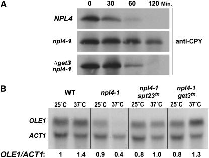
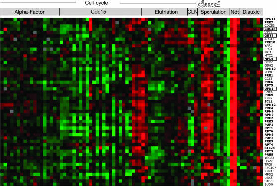
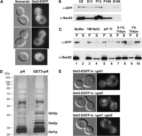
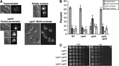
The regulation of cellular membrane dynamics is crucial for maintaining proper cell growth and division. The Cdc48-Npl4-Ufd1 complex is required for several regulated membrane-associated processes as part of the ubiquitin-proteasome system, including ER-associated degradation and the control of lipid composition in yeast. In this study we report the results of a genetic screen in Saccharomyces cerevisiae for extragenic suppressors of a temperature-sensitive npl4 allele and the subsequent analysis of one suppressor, GET3/ARR4. The GET3 gene encodes an ATPase with homology to the regulatory component of the bacterial arsenic pump. Mutants of GET3 rescue several phenotypes of the npl4 mutant and transcription of GET3 is coregulated with the proteasome, illustrating a functional relationship between GET3 and NPL4 in the ubiquitin-proteasome system. We have further found that Get3 biochemically interacts with the trans-membrane domain proteins Get1/Mdm39 and Get2/Rmd7 and that Deltaget3 is able to suppress phenotypes of get1 and get2 mutants, including sporulation defects. In combination, our characterization of GET3 genetic and biochemical interactions with NPL4, GET1, and GET2 implicates Get3 in multiple membrane-dependent pathways.
Figures

Similar articles
-
A conserved protein with AN1 zinc finger and ubiquitin-like domains modulates Cdc48 (p97) function in the ubiquitin-proteasome pathway.J Biol Chem. 2013 Nov 22;288(47):33682-33696. doi: 10.1074/jbc.M113.521088. Epub 2013 Oct 11. J Biol Chem. 2013. PMID: 24121501 Free PMC article.
-
The mechanism of tail-anchored protein insertion into the ER membrane.Mol Cell. 2011 Sep 2;43(5):738-50. doi: 10.1016/j.molcel.2011.07.020. Epub 2011 Aug 11. Mol Cell. 2011. PMID: 21835666 Free PMC article.
-
The AAA ATPase Cdc48/p97 and its partners transport proteins from the ER into the cytosol.Nature. 2001 Dec 6;414(6864):652-6. doi: 10.1038/414652a. Nature. 2001. PMID: 11740563
-
Cdc48-Ufd1-Npl4: stuck in the middle with Ub.Curr Biol. 2002 May 14;12(10):R366-71. doi: 10.1016/s0960-9822(02)00862-x. Curr Biol. 2002. PMID: 12015140 Review.
-
Quality control: another player joins the ERAD cast.Curr Biol. 2005 Dec 6;15(23):R963-4. doi: 10.1016/j.cub.2005.11.013. Curr Biol. 2005. PMID: 16332527 Review.
Cited by
-
Regulation of chaperone effects on a yeast prion by cochaperone Sgt2.Mol Cell Biol. 2012 Dec;32(24):4960-70. doi: 10.1128/MCB.00875-12. Epub 2012 Oct 8. Mol Cell Biol. 2012. PMID: 23045389 Free PMC article.
-
The ArsD As(III) metallochaperone.Biometals. 2011 Jun;24(3):391-9. doi: 10.1007/s10534-010-9398-x. Epub 2010 Dec 25. Biometals. 2011. PMID: 21188475 Free PMC article.
-
Membrane protein insertion at the endoplasmic reticulum.Annu Rev Cell Dev Biol. 2011;27:25-56. doi: 10.1146/annurev-cellbio-092910-154125. Epub 2011 Jul 21. Annu Rev Cell Dev Biol. 2011. PMID: 21801011 Free PMC article. Review.
-
The ATPase activity of Asna1/TRC40 is required for pancreatic progenitor cell survival.Development. 2018 Jan 3;145(1):dev154468. doi: 10.1242/dev.154468. Development. 2018. PMID: 29180572 Free PMC article.
-
A directed RNAi screen based on larval growth arrest reveals new modifiers of C. elegans insulin signaling.PLoS One. 2012;7(4):e34507. doi: 10.1371/journal.pone.0034507. Epub 2012 Apr 12. PLoS One. 2012. PMID: 22511947 Free PMC article.
References
-
- Adams, A., D. E. Gottschling, C. A. Kaiser and T. Stearns, 1997. Methods in Yeast Genetics. Cold Spring Harbor Laboratory Press, Plainview, NY.
-
- Albertson, R., B. Riggs and W. Sullivan, 2005. Membrane traffic: a driving force in cytokinesis. Trends Cell Biol. 15: 92–101. - PubMed
-
- Auld, K. L., C. R. Brown, J. M. Casolari, S. Komili and P. A. Silver, 2006. Genomic association of the proteasome demonstrates overlapping gene regulatory activity with transcription factor substrates. Mol. Cell 21: 861–871. - PubMed
Publication types
MeSH terms
Substances
LinkOut - more resources
Full Text Sources
Molecular Biology Databases
Miscellaneous
