. 2006 Oct;174(2):555-73.
doi: 10.1534/genetics.104.036905. Epub 2006 Jul 2.

The absence of Top3 reveals an interaction between the Sgs1 and Pif1 DNA helicases in Saccharomyces cerevisiae

Affiliations

The absence of Top3 reveals an interaction between the Sgs1 and Pif1 DNA helicases in Saccharomyces cerevisiae

Marisa Wagner et al. Genetics. 2006 Oct.

Abstract

RecQ DNA helicases and Topo III topoisomerases have conserved genetic, physical, and functional interactions that are consistent with a model in which RecQ creates a recombination-dependent substrate that is resolved by Topo III. The phenotype associated with Topo III loss suggests that accumulation of a RecQ-created substrate is detrimental. In yeast, mutation of the TOP3 gene encoding Topo III causes pleiotropic defects that are suppressed by deletion of the RecQ homolog Sgs1. We searched for gene dosage suppressors of top3 and identified Pif1, a DNA helicase that acts with polarity opposite to that of Sgs1. Pif1 overexpression suppresses multiple top3 defects, but exacerbates sgs1 and sgs1 top3 defects. Furthermore, Pif1 helicase activity is essential in the absence of Top3 in an Sgs1-dependent manner. These data clearly demonstrate that Pif1 helicase activity is required to counteract Sgs1 helicase activity that has become uncoupled from Top3. Pif1 genetic interactions with the Sgs1-Top3 pathway are dependent upon homologous recombination. We also find that Pif1 is recruited to DNA repair foci and that the frequency of these foci is significantly increased in top3 mutants. Our results support a model in which Pif1 has a direct role in the prevention or repair of Sgs1-induced DNA damage that accumulates in top3 mutants.


Figures

F<sc>igure</sc> 1.—
Figure 1.—
Pif1 is a multicopy suppressor of top3 mutant defects. (A) Overlapping nonidentical clones of PIF1 were identified in screens for high-copy suppressors of top3 slow growth. Clones 1 and 2 encompass 148,448–152,810 bp and 146,813–152,060 bp from chromosome XIII, respectively. Removal of the 1277-bp SacII (S) fragment from clone 1 that encompasses the C-terminal half of Pif1 eliminates suppression. (B) High-copy Pif1 suppresses top3 slow growth. The plate shows colony streaks of a top3Δ haploid transformed with the indicated vectors. (C) The graph indicates the doubling times at 30° for wild-type and top3Δ strains harboring the indicated vectors. Strains: wild type (W1588-4C), top3Δ (W1958 haploid segregant). Plasmids: p2μ (YEp51B), p2μ-TOP3 (HCS2), p2μ-PIF1 (clone 1).
F<sc>igure</sc> 2.—
Figure 2.—
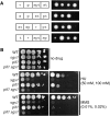
Effect of Pif1 overexpression on HU and MMS sensitivities. Shown are spot assays of 10-fold serial dilutions of wild-type and null mutant strains harboring the indicated plasmids. In each case the most concentrated spot represents 5000 cells. At low concentrations of HU (A) and MMS (B) p2μ-PIF1 partially suppresses top3Δ drug sensitivity. At higher concentrations of HU (C) and MMS (D) p2μ-PIF1 does not suppress top3Δ drug sensitivity and exacerbates sgs1Δ and sgs1Δ top3Δ drug sensitivities. Strains: wild type is W1588-4C; top3Δ, sgs1Δ, and sgs1Δ top3Δ are haploid segregants of W1588-4C. Plasmids: p2μ (YEp51B), p2μ-TOP3 (HCS2), p2μ-PIF1 (clone 1).
F<sc>igure</sc> 3.—
Figure 3.—
Toxicity caused by overexpression of the catalytically inactive Top3 protein is suppressed by overproduction of Pif1. Pictured are galactose plates on which wild-type strains (W1588-4C) cotransformed with the indicated plasmids have been streaked (see materials and methods). The empty vector controls are designated pGal. Galactose-induced expression of top3–Y356F but not TOP3 is toxic. pGal-PIF1 strongly suppresses pGal-top3-YF toxicity. In contrast, co-expression of pGal-pif1–KR does not suppress and instead exacerbates pGal-top3-YF toxicity. Top3 plasmid series is HIS3-marked: pGal (pWJ1345), pGal-TOP3 (pWJ1346), pGal-top3-YF (pWJ1347). Pif1 plasmid series is URA3-marked: pGal (pWJ1047), pGal-PIF1 (pWJ1279), pGal-pif1-KR (pWJ1280).
F<sc>igure</sc> 4.—
Figure 4.—
pif1 top3 is synthetic lethal in an Sgs1-dependent manner. (A) Four representative tetrads segregated from a sgs1Δ pif1Δ top3Δ heterozygous diploid (W3642) are shown. The genotype of each segregant is indicated: wild type (+), pif1Δ (p), sgs1Δ (s), top3Δ (t). The dead pif1Δ top3Δ segregants (pt) appear as microcolonies and do not grow further upon restreaking of the spore colony. (B) HU and MMS sensitivity of wild-type (W1588-4C) and pif1Δ, sgs1Δ, and sgs1Δ pif1Δ segregants (W3642). Tenfold serial dilutions were spotted on YPD or YPD plates containing 50 mm HU (left), 100 mm HU (right), 0.01% MMS (left), or 0.02% MMS (right). In each case, the most concentrated spot represents 50,000 cells.
F<sc>igure</sc> 5.—
Figure 5.—
The effect of Pif1 overexpression in different sgs1 allelic backgrounds. (A) The indicated sgs1 strains containing either empty vector (p2μ) or high-copy Pif1 (p2μ-PIF1) were streaked on SC-Leu plates and incubated for 3 days. (B) Wild-type and sgs1 mutants containing p2μ or p2μ-PIF1 were tested for sensitivity to HU and MMS. Shown are 10-fold serial dilutions on SC-Leu containing the indicated concentration of drug. The most concentrated spot in each case represents 50,000 cells. Strains: wild type (W1588-4C), sgs1Δ (W1958 segregant), sgs1N82 (W2069-2B), sgs1-KR (W1911-1B), sgs1-ΔN82, (W2075-3C). Plasmids: p2μ (YEp51B), p2μ-PIF1 (Clone 1).
F<sc>igure</sc> 6.—
Figure 6.—
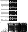
The interaction of pif1Δ with different sgs1 alleles. (A) pif1Δ top3Δ synthetic lethality is suppressed by the sgs1-K706R catalytic inactive allele. Four representative tetrads from segregation of a sgs1-K706R pif1Δ top3Δ heterozygote (W5497) are shown: wild type (+), sgs1-K706R (KR), pif1Δ (p), top3Δ (t). (B) Deletion of the Top3 interaction domain in Sgs1 (sgs1-ΔN82) is synthetic lethal with pif1Δ. Two representative tetratypes from a sgs1-ΔN82 pif1Δ heterozygote (W5493) are shown: wild type (+), sgs1-ΔN82 (N), pif1Δ (p). (C) sgs1-ΔN82 pif1Δ synthetic lethality is suppressed by the elimination of Sgs1 catalytic activity (sgs1-ΔN82, K706R). Two representative tetratypes from a sgs1-ΔN82, K706 pif1Δ heterozygote (W5495) are shown: wild type (+), sgs1-ΔN82, K706R (NKR), pif1Δ (p). (D) MMS sensitivity of viable segregants from the above crosses. Tenfold serial dilutions were spotted on SC-Leu plates with or without drug. The most concentrated spot in each case has 50,000 cells. Strains: wild type (W1588-4C), pif1Δ (W5388 segregant), sgs1Δ (W1958 segregant), sgs1Δ pif1Δ (W5489 segregant), sgs1-KR (W5491 segregant), sgs1-KR pif1Δ (W5491 segregant), sgs1-ΔN82, KR (W5495 segregant), sgs1-ΔN82, KR pif1Δ (W5495 segregant).
F<sc>igure</sc> 7.—
Figure 7.—
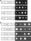
Eliminating homologous recombination suppresses pif1 top3 synthetic lethality. (A) rad54 deletion suppresses pif1 top3 lethality. Four representative tetrads from the segregation of a rad54Δ pif1Δ top3Δ heterozygous diploid (W3688) are shown: wild type (+), rad54Δ (r), pif1Δ (p), top3Δ (t). (B) rad55 deletion suppresses pif1 top3 lethality. Four representative tetrads from the segregation of a rad55Δ pif1Δ top3Δ heterozygous diploid (W3689) are shown: wild type (+), rad55Δ (r), pif1Δ (p), top3Δ (t). Similar results were obtained with rad51 and rad52 deletions (not shown).
F<sc>igure</sc> 8.—
Figure 8.—
Cellular localization of Pif1. The Pif1 protein is tagged with YFP via integration of the YFP epitope at the endogenous PIF1 locus. In all cases a Z-stack of 13 sections was taken such that the entire volume of each cell was analyzed. In A–E, the section that best emphasizes what is intended is pictured. (A) Pif1 is found in mitochondria as evidenced by colocalization with the mitochondrial marker Cox4. For reference, the position of the nucleus is indicated by Nup49, a nuclear membrane protein. Strain: W4180-8D (Pif1YFP). Plasmids: pWJ1326 (Cox4–DsRed) and pWJ1348 (CFP–Nup49). (B) Pif1 is found in the nucleus with a pronounced concentration in the nucleolus as evidenced by colocalization with the nuclear marker Nup49 and nucleolar marker Nop1. Strain: W4180-8D (Pif1YFP). Plasmids: pWJ1323 (Nup49–CFP) and pWJ1322 (Nop1–DsRed). (C–E) Pif1 occasionally localizes to discrete nuclear foci that correspond to Rad52 DNA repair foci. These Pif1 foci are induced by gamma irradiation and found with increased incidence in top3 mutant cells. Pif1 and Rad52 colocalization is shown in wild-type cells (C), in wild-type cells 1-hr after 4 krad of gamma rays (D) and in top3Δ cells (E). Strains: W4240-25B (PIF1–YFP RAD52–CFP) and haploid segregants of W4238 (top3Δ PIF1–YFP RAD52–CFP).
F<sc>igure</sc> 9.—
Figure 9.—
A model for the genetic interaction between Pif1 and Sgs1–Top3. Pif1 interacts with the Sgs1–Top3 pathway downstream of homologous recombination. A concerted Sgs1–Top3 activity is proposed to be one means of resolving recombination intermediates at the end of DNA replication, such as the double Holliday junction that is pictured (Heyer et al. 2003). In this Sgs1–Top3 resolvase model, Sgs1 molecules act on opposing DNA strands to convergently branch migrate the Holliday junctions, forming a hemicatenated strand interlink that is resolved by Top3 (middle). In the absence of Top3, the Sgs1-created substrate persists, is not efficiently resolved by other pathways, and is toxic to the cell as evidenced by the myriad defects of top3 mutants (shaded box). In the absence of Sgs1 (sgs1 or sgs1 top3 strains), this toxic substrate is never created, leaving the recombination intermediate accessible to alternative pathways (white box). Our results demonstrate that Pif1 helicase activity is required to counteract Sgs1 helicase activity that has become uncoupled from Top3. Our data suggest that Pif1 either reverses or prevents formation of this detrimental Sgs1-created DNA structure.

Similar articles

Cited by

References

    1. Akada, R., J. Yamamoto and I. Yamashita, 1997. Screening and identification of yeast sequences that cause growth inhibition when overexpressed. Mol. Gen. Genet. 254: 267–274. - PubMed
    1. Aravind, L., D. R. Walker and E. V. Koonin, 1999. Conserved domains in DNA repair proteins and evolution of repair systems. Nucleic Acids Res. 27: 1223–1242. - PMC - PubMed
    1. Bachrati, C. Z., and I. D. Hickson, 2003. RecQ helicases: suppressors of tumorigenesis and premature aging. Biochem. J. 374: 577–606. - PMC - PubMed
    1. Bennett, R. J., J. L. Keck and J. C. Wang, 1999. Binding specificity determines polarity of DNA unwinding by the Sgs1 protein of Saccharomyces cerevisiae. J. Mol. Biol. 289: 235–248. - PubMed
    1. Bennett, R. J., M. F. Noirot-Gros and J. C. Wang, 2000. Interaction between yeast sgs1 helicase and DNA topoisomerase III. J. Biol. Chem. 275: 26898–26905. - PubMed

Publication types

MeSH terms