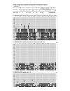
Abnormal X: autosome ratio, but normal X chromosome inactivation in human triploid cultures
- PMID: 16817970
- PMCID: PMC1526452
- DOI: 10.1186/1471-2156-7-41
Abnormal X: autosome ratio, but normal X chromosome inactivation in human triploid cultures
Abstract
Background: X chromosome inactivation (XCI) is that aspect of mammalian dosage compensation that brings about equivalence of X-linked gene expression between females and males by inactivating one of the two X chromosomes (Xi) in normal female cells, leaving them with a single active X (Xa) as in male cells. In cells with more than two X's, but a diploid autosomal complement, all X's but one, Xa, are inactivated. This phenomenon is commonly thought to suggest 1) that normal development requires a ratio of one Xa per diploid autosomal set, and 2) that an early event in XCI is the marking of one X to be active, with remaining X's becoming inactivated by default.
Results: Triploids provide a test of these ideas because the ratio of one Xa per diploid autosomal set cannot be achieved, yet this abnormal ratio should not necessarily affect the one-Xa choice mechanism for XCI. Previous studies of XCI patterns in murine triploids support the single-Xa model, but human triploids mostly have two-Xa cells, whether they are XXX or XXY. The XCI patterns we observe in fibroblast cultures from different XXX human triploids suggest that the two-Xa pattern of XCI is selected for, and may have resulted from rare segregation errors or Xi reactivation.
Conclusion: The initial X inactivation pattern in human triploids, therefore, is likely to resemble the pattern that predominates in murine triploids, i.e., a single Xa, with the remaining X's inactive. Furthermore, our studies of XIST RNA accumulation and promoter methylation suggest that the basic features of XCI are normal in triploids despite the abnormal X:autosome ratio.
Figures


Similar articles
-
The probability to initiate X chromosome inactivation is determined by the X to autosomal ratio and X chromosome specific allelic properties.PLoS One. 2009;4(5):e5616. doi: 10.1371/journal.pone.0005616. Epub 2009 May 19. PLoS One. 2009. PMID: 19440388 Free PMC article.
-
Skewed X chromosome inactivation in diploid and triploid female human embryonic stem cells.Hum Reprod. 2009 Aug;24(8):1834-43. doi: 10.1093/humrep/dep126. Epub 2009 May 8. Hum Reprod. 2009. PMID: 19429659
-
Dosage regulation of the active X chromosome in human triploid cells.PLoS Genet. 2009 Dec;5(12):e1000751. doi: 10.1371/journal.pgen.1000751. Epub 2009 Dec 4. PLoS Genet. 2009. PMID: 19997486 Free PMC article.
-
Female-bias in systemic lupus erythematosus: How much is the X chromosome to blame?Biol Sex Differ. 2024 Oct 7;15(1):76. doi: 10.1186/s13293-024-00650-y. Biol Sex Differ. 2024. PMID: 39375734 Free PMC article. Review.
-
X-inactive-specific transcript: a long noncoding RNA with a complex role in sex differences in human disease.Biol Sex Differ. 2024 Dec 5;15(1):101. doi: 10.1186/s13293-024-00681-5. Biol Sex Differ. 2024. PMID: 39639337 Free PMC article. Review.
Cited by
-
Dosage compensation of the sex chromosomes.Annu Rev Genet. 2012;46:537-60. doi: 10.1146/annurev-genet-110711-155454. Epub 2012 Sep 4. Annu Rev Genet. 2012. PMID: 22974302 Free PMC article. Review.
-
Silencing XIST on the future active X: Searching human and bovine preimplantation embryos for the repressor.Eur J Hum Genet. 2024 Apr;32(4):399-406. doi: 10.1038/s41431-022-01115-9. Epub 2022 May 19. Eur J Hum Genet. 2024. PMID: 35585273 Free PMC article.
-
Ovarian follicles of young patients with Turner's syndrome contain normal oocytes but monosomic 45,X granulosa cells.Hum Reprod. 2019 Sep 29;34(9):1686-1696. doi: 10.1093/humrep/dez135. Hum Reprod. 2019. PMID: 31398245 Free PMC article. Clinical Trial.
-
A self-enhanced transport mechanism through long noncoding RNAs for X chromosome inactivation.Sci Rep. 2016 Aug 16;6:31517. doi: 10.1038/srep31517. Sci Rep. 2016. PMID: 27527711 Free PMC article.
-
A new model for random X chromosome inactivation.Development. 2009 Jan;136(1):1-10. doi: 10.1242/dev.025908. Epub 2008 Nov 26. Development. 2009. PMID: 19036804 Free PMC article.
References
Publication types
MeSH terms
Substances
Grants and funding
LinkOut - more resources
Full Text Sources

