

doi: 10.1101/gad.1430906.
A positive feedback loop couples Ras activation and CD44 alternative splicing
Affiliations
- PMID: 16818603
- PMCID: PMC1522067
- DOI: 10.1101/gad.1430906
Item in Clipboard
A positive feedback loop couples Ras activation and CD44 alternative splicing
Genes Dev.
.
Abstract
The Ras signaling pathway is important in both cell proliferation and tumor progression. Alternatively spliced isoforms of CD44 containing variable exon 6 (v6) can serve as coreceptors for growth factor receptors that activate Ras. Here we use v6-specific small interfering RNA (siRNA) to investigate the role of CD44 alternative splicing in Ras signaling. We identify a positive feedback loop in which Ras signaling promotes CD44v6 splicing, and CD44v6 then sustains late Ras signaling, which is important for cell cycle progression. These results are the first demonstration of a positive feedback loop linking signaling-dependent alternative splicing to mitogenic progression.
Figures
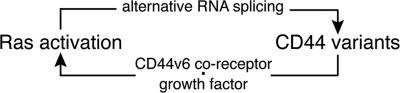
A model of a positive feedback loop between Ras activation and CD44 variants. Activated Ras signaling stimulates CD44 alternative splicing, resulting in the production of CD44 variants. Subsequently, the spliced CD44 variants containing v6 exon act as coreceptors. They form complexes with growth factors and their receptor tyrosine kinases and further activate Ras signaling.
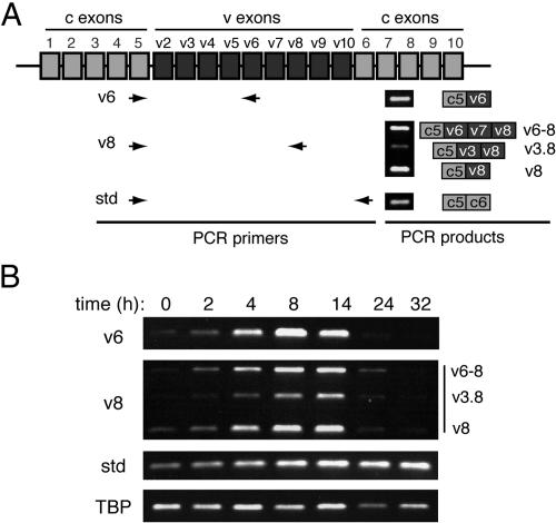
Expression of CD44 variants is stimulated by growth factors in serum. (A) A schematic diagram of the CD44 gene. Both constitutive (c) and variable (v) exons are depicted. The PCR primers used to amplify CD44 variable and standard (std) isoforms are shown as arrowheads. The primers for both the v6 and standard isoforms predominantly generate one PCR product, whereas the primers for the v8 variants amplify three splice variants. Structures of the PCR products are depicted to the right of the individual example gels. (B) A time course of serum stimulation. Serum-starved HeLa cells were stimulated with 10% serum and collected at the indicated time points. RNA was extracted, and quantitative RT–PCR was performed. Agarose gels of the PCR products are shown. The numbers of PCR cycles and exposure length of the images for each set of PCR primers vary and cannot be directly compared. PCR primers used in each reaction are depicted to the left of the gels. TATA-binding protein (TBP) served as a loading control.
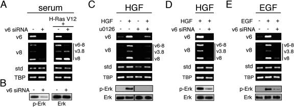
siRNA-mediated knockdown of CD44v6 affects alternative splicing of CD44 and down-regulates Erk activity. (A) RT–PCR analyses of CD44 variants in HeLa cells transfected with control siRNA or v6 siRNA followed by serum stimulation for 8 h (left panel), or transfected with an additional H-Ras V12 construct (right panel). TBP was used as a loading control. (B) Western blot analyses of Erk phosphorylation following serum stimulation for 8 h in cells transfected with control or v6 siRNA. Antibodies recognizing phosphorylated Erk (p-Erk) or total Erk (Erk) were used. (C–E) RT–PCR analyses of CD44 variants and Western blot analyses of Erk phosphorylation in serum-starved HeLa cells that were stimulated with HGF (60 ng/mL) for 8 h in the absence or presence of U0126 (25 μM) (C), or in serum starved control or v6 siRNA-treated HeLa that were stimulated with HGF (D) or EGF (5 ng/mL, E) for 8 h.
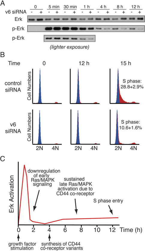
Down-regulation of CD44v6 impairs sustained Erk activation and inhibits the G1–S transition. (A) Western blot analyses of Erk activation in EGF-stimulated HeLa cells transfected with control or CD44v6 siRNA. These cells were collected at indicated times after EGF addition. (B) Cell cycle analyses of serum-starved T98G cells, treated with control or CD44v6 siRNA, and stimulated with serum to re-enter the cell cycle. The blue peak represents cells in G0/G1 phase at time 0 h. This peak is overlaid on the subsequent time points for each experiment. The percentage of cells in S phase at 15 h after serum stimulation is indicated. (C) A model for sustained Ras/MAPK signaling and G1–S transition, dependent on CD44 coreceptor variants. See text for details.
References
-
- Bourguignon L.Y., Zhu H., Chu A., Iida N., Zhang L., Hung M.C. Interaction between the adhesion receptor, CD44, and the oncogene product, p185HER2, promotes human ovarian tumor cell activation. J. Biol. Chem. 1997;272:27913–27918. - PubMed
-
- Favata M.F., Horiuchi K.Y., Manos E.J., Daulerio A.J., Stradley D.A., Feeser W.S., Van Dyk D.E., Pitts W.J., Earl R.A., Hobbs F., et al. Identification of a novel inhibitor of mitogen-activated protein kinase kinase. J. Biol. Chem. 1998;273:18623–18632. - PubMed
Publication types
MeSH terms
Substances
LinkOut - more resources
Full Text Sources
Other Literature Sources
Miscellaneous