. 2006 Jul 1;20(13):1744-54.
doi: 10.1101/gad.1435106.

Maternal BRG1 regulates zygotic genome activation in the mouse

Affiliations

Maternal BRG1 regulates zygotic genome activation in the mouse

Scott J Bultman et al. Genes Dev. .

Abstract

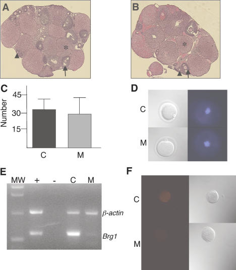
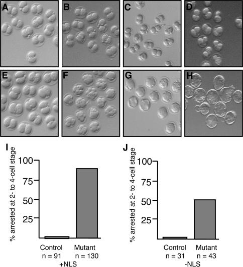
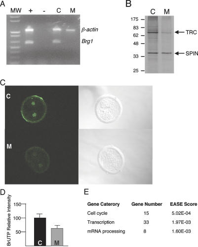
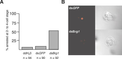
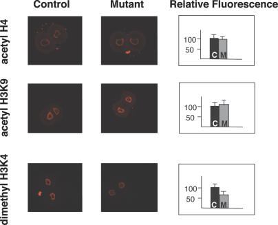
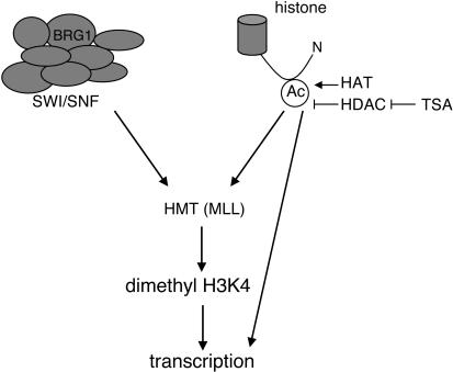
Zygotic genome activation (ZGA) is a nuclear reprogramming event that transforms the genome from transcriptional quiescence at fertilization to robust transcriptional activity shortly thereafter. The ensuing gene expression profile in the cleavage-stage embryo establishes totipotency and is required for further development. Although little is known about the molecular basis of ZGA, oocyte-derived mRNAs and proteins that alter chromatin structure are likely crucial. To test this hypothesis, we generated a maternal-effect mutation of Brg1, which encodes a catalytic subunit of SWI/SNF-related complexes, utilizing Cre-loxP gene targeting. In conditional-mutant females, BRG1-depleted oocytes completed meiosis and were fertilized. However, embryos conceived from BRG1-depleted eggs exhibited a ZGA phenotype including two-cell arrest and reduced transcription for approximately 30% of expressed genes. Genes involved in transcription, RNA processing, and cell cycle regulation were particularly affected. The early embryonic arrest is not a consequence of a defective oocyte because depleting maternal BRG1 after oocyte development is complete by RNA interference (RNAi) also resulted in two-cell arrest. To our knowledge, Brg1 is the first gene required for ZGA in mammals. Depletion of maternal BRG1 did not affect global levels of histone acetylation, whereas dimethyl-H3K4 levels were reduced. These data provide a framework for understanding the mechanism of ZGA.


Figures

Figure 1.
Figure 1.
Analysis of Brg1Zp3-Cre mutant germline. (A,B) H&E-stained ovary sections from wild-type control (A) and conditional mutant (B) females at 100× magnification. Primary follicles (arrowheads), secondary follicles (arrows) follicles, and corpora lutea (*) are indicated. (C) Average number of unfertilized eggs recovered from superovulated wild-type control (C) and conditional mutant (M) females. (D) DIC (top) and DAPI (bottom) images of wild-type control (C) and conditional mutant (M) ovulated eggs at 400× magnification. Polar bodies and MII-arrested chromosomes are visible in DIC and DAPI images, respectively. (E,F) Brg1 gene product is depleted in unfertilized eggs from conditional mutant females. (E) Image of ethidium bromide stained agarose gel containing β-actin (1020 base pairs [bp]) and Brg1 (585 bp) RT–PCR products. (MW) One-kilobase ladder molecular-weight standard; (+) positive control consisting of wild-type neonatal tissues; (−), negative control consisting of wild-type neonatal tissues except reverse transcriptase was omitted from RT reaction; (C) 20 unfertilized eggs from wild-type control; (M) 20 unfertilized eggs from conditional mutant. (F) IF of BRG1 protein in unfertilized eggs from wild-type control (C) or conditional mutant (M) females. DIC images are shown to the right.
Figure 2.
Figure 2.
Cleavage-stage arrest of Brg1Zp3-Cre maternally depleted embryos. (AH) Representative bright-field photographs from an embryo culture experiment. Maternally depleted (A) and control (E) embryos were flushed out of oviducts at the two-cell stage on E1.5. Maternally depleted (AD) and control (EH) embryos were subsequently cultured for 1 d (equivalent of E2.5) (B,F), 2 d (equivalent of E3.5) (C,G), or 3 d (equivalent of E4.5) (D,H). Maternally depleted embryos remained at the two-cell stage throughout the culture period (B–D). Control embryos advanced to the uncompacted and compacted morula stages (F), compacted morula and blastocyst stages (G), and expanded blastocyst stage (H). Embryos came from control (Brg1+/floxed, TgZp3-Cre) or Brg1Zp3-Cre females bred to wild-type males. (I) Quantification of the two- to four-cell arrest in control (C) and maternally depleted (M) embryos after the 3-d culture period. Numbers of embryos cultured are indicated. (J) Quantification of the two- to four-cell arrest in embryos with a Zp3-Cre transgene lacking a NLS after 3-d culture period. Numbers of embryos cultured are indicated.
Figure 3.
Figure 3.
Reduced gene expression in Brg1Zp3-Cre maternally depleted embryos. (A) Image of ethidium bromide-stained agarose gel containing β-actin (1020 bp) and Brg1 (585 bp) RT–PCR products. (MW) One-kilobase ladder molecular-weight standard; (+) positive control consisting of wild-type neonatal tissues; (−) negative control consisting of wild-type neonatal tissues except reverse transcriptase was omitted from RT reaction; (C) two-cell embryos from wild-type control female (n = 15); (M) maternally depleted two-cell embryos (n = 15). (B) 35S-methionine metabolic labeling of a pool of six control (C) and six maternally depleted (M) two-cell embryos. Shown is a representative autoradiograph of a polyacrylamide gel with molecular weights (in kilodaltons) indicated at the left and the TRC and SPIN proteins indicated at the right. (C) BrUTP incorporation detected by IF as a measure of genome-wide transcription. Representative confocal images of control (C, top) and maternally depleted (M, bottom) two-cell embryos are shown. (Right panels) DIC images are also shown. (D) Quantification of BrUTP incorporation data. Relative intensity of control (C) and maternally depleted (M) embryos. (E) EASE analysis of selected biological processes.
Figure 4.
Figure 4.
Perturbation of BRG1 by RNAi results in cleavage-stage arrest and reduced protein levels. (A) Quantification of two- to four-cell arrest after wild-type zygotes were injected with water (ddH2O), dsGFP RNA, or dsBrg1 RNA and cultured for 4 d. Numbers of zygotes injected are indicated. (B) Representative confocal images detecting BRG1 protein by IF in three-cell embryos injected with dsGPF (top) or dsBrg1 (bottom) RNAs. BRG1 protein is nuclear but excluded from nucleoli (dark spots). Only one nucleus is visible in optical slice shown for the dsGFP panel; the other two nuclei express similar levels of BRG1 protein as shown in Supplementary Figure S3. In contrast, all three nuclei are in optimal focal plane for dsBrg1 panel. DIC images are shown to the right.
Figure 5.
Figure 5.
Normal and aberrant covalent histone modifications. Representative confocal images detecting covalent histone modifications by IF in control (genotype of dam: floxed/floxed, no transgene) and maternally depleted (Mutant) embryos. Anti-pan-acetyl-H4 (top row), anti-acetyl-H3K9 (middle row), and anti-dimethyl-H3K4 (bottom row) primary antibodies were utilized. Signal is excluded from nucleoli (dark spots in center). The small, bright spots corresponding to neither nucleus in the top right and bottom left panels are polar bodies. Quantification of the IF data in control (C) and maternally depleted (M) embryos is shown furthest to the right under Relative Fluorescence heading.
Figure 6.
Figure 6.
A model depicting the relationship of SWI/SNF-related complexes and covalent histone modifications during ZGA. Maternally derived SWI/SNF-related complexes (left) and histone acetylation (right) converge to stimulate MLL. MLL methylates H3K4, which, in turn, results in transcription. In Brg1Zp3-Cre maternally depleted embryos, dimethyl H3K4 and transcription are reduced but not abolished, because the histone acetylation input is intact and other chromatin remodeling complexes might compensate. For the same reason, TSA treatment of maternally depleted embryos increases histone acetylation and restores dimethyl H3K4 to levels of untreated controls and transcription to higher than normal levels nearly equal to TSA-treated controls. It is also likely that histone acetylation stimulates transcription in a dimethyl H3K4-independent manner (line from Ac to transcription). (Ac) Acetyl group on N-terminal tail of a core histone; (HAT) histone acetyltransferase; (HDAC) histone deacetylase; (TSA) trichostatin A; (HMT) histone methyltransferase; (MLL) mixed lineage leukemia.

References

    1. Agalioti T., Chen G., Thanos D. Deciphering the transcriptional histone acetylation code for a human gene. Cell. 2002;111:381–392. - PubMed
    1. Aoki F., Worrad D.M., Schultz R.M. Regulation of transcriptional activity during the first and second cell cycles in the preimplantation mouse embryo. Dev. Biol. 1997;181:296–307. - PubMed
    1. Ayton P., Sneddon S.F., Palmer D.B., Rosewell I.R., Owen M.J., Young B., Presley R., Subramanian V. Truncation of the Mll gene in exon 5 by gene targeting leads to early preimplantation lethality of homozygous embryos. Genesis. 2001;30:201–212. - PubMed
    1. Bazett-Jones D.P., Cote J., Landel C.C., Peterson C.L., Workman J.L. The SWI/SNF complex creates loop domains in DNA and polynucleosome arrays and can disrupt DNA–histone contacts within these domains. Mol. Cell. Biol. 1999;19:1470–1478. - PMC - PubMed
    1. Bellotto M., Bopp D., Senti K.A., Burke R., Deak P., Maroy P., Dickson B., Basler K., Hafen E. Maternal-effect loci involved in Drosophila oogenesis and embryogenesis: P element-induced mutations on the third chromosome. Int. J. Dev. Biol. 2002;46:149–157. - PubMed

Publication types

MeSH terms