

Sustained expression of early growth response protein-1 blocks angiogenesis and tumor growth
- PMID: 16818645
- PMCID: PMC2882226
- DOI: 10.1158/0008-5472.CAN-05-2732
Sustained expression of early growth response protein-1 blocks angiogenesis and tumor growth
Abstract
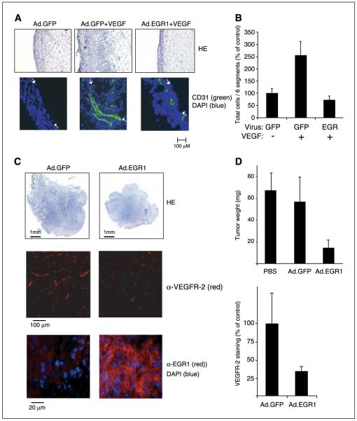
Transient induction of the transcription factor early growth response protein-1 (EGR-1) plays a pivotal role in the transcriptional response of endothelial cells to the angiogenic growth factors vascular endothelial growth factor (VEGF) and basic fibroblast growth factor (bFGF), which are produced by most tumors and are involved in the angiogenic switch. We report here that sustained expression of EGR-1 by recombinant adenoviruses in endothelial cells, however, leads to the specific induction of potent feedback inhibitory mechanisms, including strong up-regulation of transcriptional repressors, negative cell cycle check point effectors, proteins with established antiangiogenic activity, and several proapoptotic genes. Sustained EGR-1 expression consistently leads to an antiangiogenic state characterized by an altered responsiveness to VEGF and bFGF and a striking inhibition of sprouting and tubule formation in vitro. Furthermore, EGR-1-expressing viruses potently inhibit cell invasion and vessel formation in the murine Matrigel model and repress tumor growth in a murine fibrosarcoma model. We propose that gene therapy involving sustained EGR-1 expression may constitute a novel therapeutic principle in the treatment of cancer due to the simultaneous induction of multiple pathways of antiangiogenesis, growth arrest, and apoptosis induction in proliferating cells leading to preferential inhibition of angiogenesis and tumor growth.
Figures


Similar articles
-
Fibulins 3 and 5 antagonize tumor angiogenesis in vivo.Cancer Res. 2006 Mar 1;66(5):2621-9. doi: 10.1158/0008-5472.CAN-04-4096. Cancer Res. 2006. PMID: 16510581
-
Vascular endothelial growth factor activation of endothelial cells is mediated by early growth response-3.Blood. 2010 Mar 25;115(12):2520-32. doi: 10.1182/blood-2009-07-233478. Epub 2009 Nov 23. Blood. 2010. PMID: 19965691 Free PMC article.
-
NAB2, a corepressor of EGR-1, inhibits vascular endothelial growth factor-mediated gene induction and angiogenic responses of endothelial cells.J Biol Chem. 2003 Mar 28;278(13):11433-40. doi: 10.1074/jbc.M204937200. Epub 2002 Nov 8. J Biol Chem. 2003. PMID: 12427750
-
Early growth response-1: blocking angiogenesis by shooting the messenger.Cell Cycle. 2004 Jan;3(1):10-1. Cell Cycle. 2004. PMID: 14657654 Review.
-
Targeting Angiogenesis in Cancer Therapy: Moving Beyond Vascular Endothelial Growth Factor.Oncologist. 2015 Jun;20(6):660-73. doi: 10.1634/theoncologist.2014-0465. Epub 2015 May 22. Oncologist. 2015. PMID: 26001391 Free PMC article. Review.
Cited by
-
FOXO3 modulates endothelial gene expression and function by classical and alternative mechanisms.J Biol Chem. 2010 Apr 2;285(14):10163-78. doi: 10.1074/jbc.M109.056663. Epub 2010 Feb 1. J Biol Chem. 2010. PMID: 20123982 Free PMC article.
-
Egr-1 upregulates Siva-1 expression and induces cardiac fibroblast apoptosis.Int J Mol Sci. 2014 Jan 21;15(1):1538-53. doi: 10.3390/ijms15011538. Int J Mol Sci. 2014. PMID: 24451137 Free PMC article.
-
Dysregulation of CXCL9 and reduced tumor growth in Egr-1 deficient mice.J Hematol Oncol. 2009 Feb 7;2:7. doi: 10.1186/1756-8722-2-7. J Hematol Oncol. 2009. PMID: 19200397 Free PMC article.
-
NGFI-A Binding Protein 2 Promotes EGF-Dependent HNSCC Cell Invasion.Cancers (Basel). 2019 Mar 6;11(3):315. doi: 10.3390/cancers11030315. Cancers (Basel). 2019. PMID: 30845713 Free PMC article.
-
Egr-1 activation by cancer-derived extracellular vesicles promotes endothelial cell migration via ERK1/2 and JNK signaling pathways.PLoS One. 2014 Dec 12;9(12):e115170. doi: 10.1371/journal.pone.0115170. eCollection 2014. PLoS One. 2014. PMID: 25502753 Free PMC article.
References
-
- Folkman J, Kalluri R. Cancer without disease. Nature. 2004;427:787. - PubMed
-
- Carmeliet P. Manipulating angiogenesis in medicine. J Intern Med. 2004;255:538–61. - PubMed
-
- Ferrara N. Vascular endothelial growth factor: basic science and clinical progress. Endocr Rev. 2004;25:581–611. - PubMed
-
- Gashler A, Sukhatme VP. Early growth response protein 1 (Egr-1): prototype of a zinc-finger family of transcription factors. Prog Nucleic Acid Res Mol Biol. 1995;50:191–224. - PubMed
-
- Fahmy RG, Dass CR, Sun LQ, Chesterman CN, Khachigian LM. Transcription factor Egr-1 supports FGF-dependent angiogenesis during neovascularization and tumor growth. Nat Med. 2003;9:1026–32. - PubMed
Publication types
MeSH terms
Substances
Grants and funding
LinkOut - more resources
Full Text Sources
Other Literature Sources
Molecular Biology Databases
Research Materials
