Role of HMGB1 in apoptosis-mediated sepsis lethality
- PMID: 16818669
- PMCID: PMC2118346
- DOI: 10.1084/jem.20052203
Role of HMGB1 in apoptosis-mediated sepsis lethality
Abstract
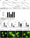
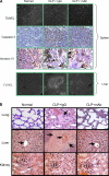
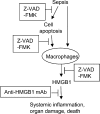
Severe sepsis, a lethal syndrome after infection or injury, is the third leading cause of mortality in the United States. The pathogenesis of severe sepsis is characterized by organ damage and accumulation of apoptotic lymphocytes in the spleen, thymus, and other organs. To examine the potential causal relationships of apoptosis to organ damage, we administered Z-VAD-FMK, a broad-spectrum caspase inhibitor, to mice with sepsis. We found that Z-VAD-FMK-treated septic mice had decreased levels of high mobility group box 1 (HMGB1), a critical cytokine mediator of organ damage in severe sepsis, and suppressed apoptosis in the spleen and thymus. In vitro, apoptotic cells activate macrophages to release HMGB1. Monoclonal antibodies against HMGB1 conferred protection against organ damage but did not prevent the accumulation of apoptotic cells in the spleen. Thus, our data indicate that HMGB1 production is downstream of apoptosis on the final common pathway to organ damage in severe sepsis.
Figures


Similar articles
-
Splenectomy protects against sepsis lethality and reduces serum HMGB1 levels.J Immunol. 2008 Sep 1;181(5):3535-9. doi: 10.4049/jimmunol.181.5.3535. J Immunol. 2008. PMID: 18714026 Free PMC article.
-
The release of high mobility group box 1 in apoptosis is triggered by nucleosomal DNA fragmentation.Arch Biochem Biophys. 2011 Feb 15;506(2):188-93. doi: 10.1016/j.abb.2010.11.011. Epub 2010 Nov 17. Arch Biochem Biophys. 2011. PMID: 21093407
-
Local thymic caspase-9 inhibition improves survival during polymicrobial sepsis in mice.J Mol Med (Berl). 2006 May;84(5):389-95. doi: 10.1007/s00109-005-0017-1. Epub 2006 Feb 2. J Mol Med (Berl). 2006. PMID: 16453149
-
HMGB1 as a DNA-binding cytokine.J Leukoc Biol. 2002 Dec;72(6):1084-91. J Leukoc Biol. 2002. PMID: 12488489 Review.
-
The role of high mobility group box-1 protein in severe sepsis.Curr Opin Infect Dis. 2006 Jun;19(3):231-6. doi: 10.1097/01.qco.0000224816.96986.67. Curr Opin Infect Dis. 2006. PMID: 16645483 Review.
Cited by
-
Gastric alarmin release: A warning signal in the development of gastric mucosal diseases.Front Immunol. 2022 Oct 6;13:1008047. doi: 10.3389/fimmu.2022.1008047. eCollection 2022. Front Immunol. 2022. PMID: 36275647 Free PMC article. Review.
-
Secretory autophagy machinery and vesicular trafficking are involved in HMGB1 secretion.Autophagy. 2021 Sep;17(9):2345-2362. doi: 10.1080/15548627.2020.1826690. Epub 2020 Oct 5. Autophagy. 2021. PMID: 33017561 Free PMC article.
-
A potential new pathway for heparin treatment of sepsis-induced lung injury: inhibition of pulmonary endothelial cell pyroptosis by blocking hMGB1-LPS-induced caspase-11 activation.Front Cell Infect Microbiol. 2022 Sep 15;12:984835. doi: 10.3389/fcimb.2022.984835. eCollection 2022. Front Cell Infect Microbiol. 2022. PMID: 36189354 Free PMC article. Review.
-
Bench-to-bedside review: High-mobility group box 1 and critical illness.Crit Care. 2007;11(5):229. doi: 10.1186/cc6088. Crit Care. 2007. PMID: 17903310 Free PMC article.
-
Transcriptomic and ultrastructural evidence indicate that anti-HMGB1 antibodies rescue organic dust-induced mitochondrial dysfunction.Cell Tissue Res. 2022 May;388(2):373-398. doi: 10.1007/s00441-022-03602-3. Epub 2022 Mar 4. Cell Tissue Res. 2022. PMID: 35244775 Free PMC article.
References
-
- Angus, D., and R.S. Wax. 2001. Epidemiology of sepsis: an update. Crit. Care Med. 29:S109–S116. - PubMed
-
- Riedemann, N.C., R.-F. Guo, and P.A. Ward. 2003. Novel strategies for the treatment of sepsis. Nat. Med. 9:517–524. - PubMed
-
- Tracey, K.J. 2005. Fatal Sequence: the Killer Within. Dana Press, Washington, D.C. 224 pp.
-
- Lotze, M.T., and K.J. Tracey. 2005. High-mobility group box 1 protein (HMGB1): nuclear weapon in the immune arsenal. Nat. Rev. Immunol. 5:331–342. - PubMed
-
- Hotchkiss, R., K.C. Chang, P.E. Swanson, K.W. Tinsley, J.J. Hui, P. Klender, S. Xanthoudakis, S. Roy, C. Black, E. Grimm, et al. 2000. Caspase inhibitors improve survival in sepsis: a critical role of the lymphocyte. Nat. Immunol. 1:496–501. - PubMed
Publication types
MeSH terms
Substances
Grants and funding
LinkOut - more resources
Full Text Sources
Other Literature Sources
Medical

