Case Reports
. 2006 Jul 10;203(7):1745-59.
doi: 10.1084/jem.20060085. Epub 2006 Jul 3.

X-linked susceptibility to mycobacteria is caused by mutations in NEMO impairing CD40-dependent IL-12 production

Affiliations
Case Reports

X-linked susceptibility to mycobacteria is caused by mutations in NEMO impairing CD40-dependent IL-12 production

Orchidée Filipe-Santos et al. J Exp Med. .

Abstract

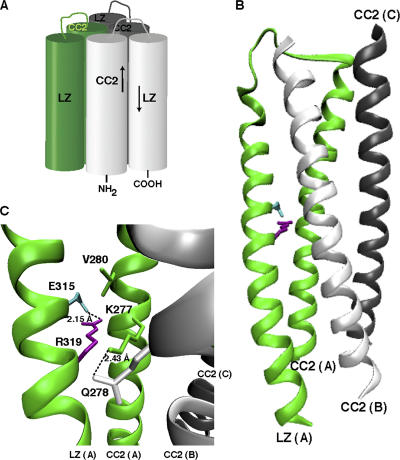
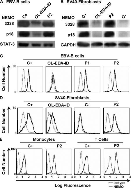
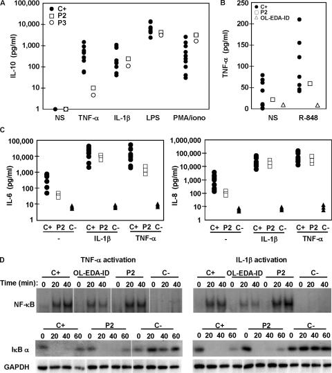
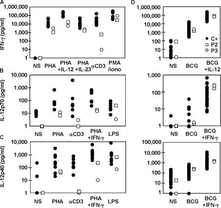
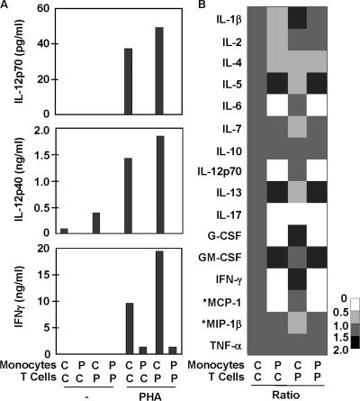
Germline mutations in five autosomal genes involved in interleukin (IL)-12-dependent, interferon (IFN)-gamma-mediated immunity cause Mendelian susceptibility to mycobacterial diseases (MSMD). The molecular basis of X-linked recessive (XR)-MSMD remains unknown. We report here mutations in the leucine zipper (LZ) domain of the NF-kappaB essential modulator (NEMO) gene in three unrelated kindreds with XR-MSMD. The mutant proteins were produced in normal amounts in blood and fibroblastic cells. However, the patients' monocytes presented an intrinsic defect in T cell-dependent IL-12 production, resulting in defective IFN-gamma secretion by T cells. IL-12 production was also impaired as the result of a specific defect in NEMO- and NF-kappaB/c-Rel-mediated CD40 signaling after the stimulation of monocytes and dendritic cells by CD40L-expressing T cells and fibroblasts, respectively. However, the CD40-dependent up-regulation of costimulatory molecules of dendritic cells and the proliferation and immunoglobulin class switch of B cells were normal. Moreover, the patients' blood and fibroblastic cells responded to other NF-kappaB activators, such as tumor necrosis factor-alpha, IL-1beta, and lipopolysaccharide. These two mutations in the NEMO LZ domain provide the first genetic etiology of XR-MSMD. They also demonstrate the importance of the T cell- and CD40L-triggered, CD40-, and NEMO/NF-kappaB/c-Rel-mediated induction of IL-12 by monocyte-derived cells for protective immunity to mycobacteria in humans.


Figures

Figure 1.
Figure 1.
Clinical phenotypes and NEMO genotypes of patients. (A) Pedigrees of three kindreds with NEMO mutations (A, B, and C). Each generation is designated by a Roman numeral, and each individual is designated by an Arabic numeral. Male patients (in black squares) present mycobacterial infections and hemizygous NEMO mutations; asymptomatic female carriers are represented by black dots. The probands are indicated by arrows. (B) The teeth of patient P1 (Kindred A, II.4, sparse teeth), patient P2 (Kindred B, II.1, conical decidual incisors), and patient P3 (Kindred C, II.1, normal teeth). (C) Sequence electrophoregram of NEMO complementary DNA in the region corresponding to the mutation. Kindred A, P1 presents the mutation A→C leading to the replacement at residue 315 of Glu (E) by Ala (A) (E315A). Kindred B, P2 and kindred C, P3 present the mutation G→A resulting in the replacement of an Arg (R) at position 319 by Gln (Q) (R319Q). (D) Schematic representation of the NEMO coding region, from exons 2 to 10, and corresponding domains, shown in dark gray: the coil-coiled domain 1 (CC1, Leu 93 to Gln 194) and coil-coiled domain 2 (CC2, Met 258 to Lys 292), and the leucine zipper (LZ, Tyr 308 to Lys 344) and zinc finger (ZF, Cys 397 to Cys 417) domains. All published hypomorphic NEMO mutations (associated with the various forms of EDA-ID) are represented (reference 43). Missense and nonsense mutations (amino-acid code) are shown on top, whereas splice and frameshift mutations (nucleotide code) are shown on the bottom. NEMO mutations in red represent mutations associated with mycobacterial infections.
Figure 2.
Figure 2.
A model of the three-dimensional structure of NEMO. (A) Schematic representation of the structural model of the NEMO oligomerization domain. The oligomerization domains from three NEMO subunits are represented in green, light gray, and dark gray. The LZ motifs dock in an antiparallel manner in the crevices defined by the central trimeric CC2 coiled-coil. (B) Modeling of NEMO oligomerization domain. Modeling was based on the coordinates of the HIV-1 gp41 ectodomain, as described in Materials and methods. For the sake of simplicity, only one LZ domain (LZ[A], in green) is represented, together with the three CC2 motifs forming the core of the pseudo-six-helix bundle. Amino acids E315 and R319, mutated in the NEMO protein of the XR-MSMD patients and located within the LZ motif, are shown in blue and purple, respectively. (C) A close-up of the surrounding environment of E315 (blue) and R319 (purple) amino acids, which probably form an intramolecular salt bridge. Spatially close to these two residues, V280 and K277 (green) are located within the CC2 motif of the same NEMO subunit. Q278 (light gray), located within the CC2 motif of the adjacent B subunit (CC2[B]) probably forms an intermolecular hydrogen bond with K277.
Figure 3.
Figure 3.
Expression of NEMO in the patients. NEMO protein, detected by Western blotting with two specific antibodies, 3328 and p18, which recognize different parts of the N-terminus of NEMO (A) EBV-transformed B cell lines and (B) SV-40–transformed fibroblastic cell lines from a healthy donor (C+), an OL-EDA-ID patient carrying the X420W NEMO mutation, P2 and an SV-40–transformed fibroblastic cell line from a NEMO-deficient fetus as a negative control (C−). Intracellular staining of NEMO with a specific antibody recognizing amino acids 278–396 and detected by flow cytometry in (C) EBV-transformed B cell lines and (D) SV40-transformed fibroblastic cell lines from healthy control (C+), an OL-EDA-ID patient, P1 and P2. (E) Purified monocytes (left) and T cells (right) from a healthy control (C+) and P2.
Figure 4.
Figure 4.
NF-κB activation in leukocytes and fibroblasts. Healthy controls (C+, •), OL-EDA-ID patient (▵), SV-40–transformed fibroblastic cell line from NEMO-deficient fetus (C−,▴), P2 (□), and P3 (◯). (A) IL-10 production by whole blood cells from 10 healthy controls and P2 and P3, upon stimulation with LPS (1 μg/ml), TNF-α (20 ng/ml), IL-1β (10 ng/ml), and PMA/ionomycin (10−7/10−5 M) for 48 h. (B) TNF-α production by EBV-transformed B cells after 24 h of TLR7/8 activation by R-848 agonist. (C) IL-6 and IL-8 production by SV-40–transformed fibroblastic cell lines from nine healthy controls and from P2 and C− (three experiments each for P2 and C−), after 24 h of TNF-α and IL-1β stimulation. All cytokines were determined by sandwich ELISA. (D) DNA-binding activity in nuclear extracts from fibroblastic cell lines after TNF-α (20 ng/ml) and IL-1β (10 ng/ml) activation for various lengths of time, as detected with a radio-labeled DNA probe by EMSA (top), and IκBα degradation and GAPDH expression, detected by Western blotting, in the corresponding cytoplasmic extracts (bottom). The results shown are representative of at least two independent experiments.
Figure 5.
Figure 5.
IL-12 and IFN-γ production by leukocytes. Cytokine production by PBMCs from 10 healthy donors, (C+, •), P2 (□), and P3 (◯) in response to PHA, alone or in combination with recombinant IL-12 (20 ng/ml), IL-23 (20 ng/ml), or IFNγ (5,000 UI/ml), anti-CD3 (10 ng/ml), LPS (1 μg/ml) and PMA/ionomycin (10−7/10−5 M). (A) IFN-γ, (B) IL-12p70, (C) IL-12p40 production. (D) Cytokine production by whole blood cells from 50 healthy donors, P2, and P3 upon stimulation with live BCG alone or in combination with IL-12 (20 ng/ml) or IFN-γ (5,000 IU/ml): IFN-γ (top), IL-12p70 (middle), and IL-12p40 (bottom) secretion. For each patient, the results shown are representative of two independent experiments.
Figure 6.
Figure 6.
IL-12 and IFN-γ production by cocultured monocytes and T cells from the patients studied and healthy controls. (A) IL-12p70, IL-12p40, and IFN-γ production, measured using classical sandwich ELISA, in a mixture of purified monocytes and T cells, as indicated upon stimulation with PHA. The results shown are representative of three independent experiments for P2 and one for P3. (B) The same coculture supernatants were analyzed for a multiplex of 16 cytokines, using the Bioplex array. Each column represents the data for one monocyte–T cell coculture system, and all four columns correspond to the same experiment. Each row corresponds to one cytokine. The gray-scale bar indicates the magnitude of cytokine expression, using the control/control (C/C) coculture system as a reference. For each data point, the amount of cytokine produced in the unstimulated system was subtracted from that produced in the PHA-activated system, and the result obtained was compared with the reference value (C/C). The production of *MCP-1 and *MIP-1β by monocytes was PHA-dependent but T cell–independent, as monocytes responded to PHA by producing large amounts of these cytokines, whereas the addition of T cells did not increase cytokine production. The defects in the production of IL-6, IL-12p70, G-CSF, IFN-γ, and MCP-1 were confirmed in three independent experiments on blood cells from P2 and one experiment on blood from P3.
Figure 7.
Figure 7.
The impact of CD40 and CD40L deficiency on IL-12 and IFN-γ production. IL-12p70, IL-12p40, and IFN-γ production, measured by classical sandwich ELISA, in a mixture of purified monocytes and T cells, as indicated, upon stimulation with PHA from a healthy donor with (A) CD40L- and (B) CD40-deficient PBMCs. MDDCs obtained from two healthy controls (C2 and C3), P2 and P3, after 24 h of incubation alone or with LPS (1 μg/ml) plus IL-1β (10 ng/ml) as a control of CD40-independent activation, cocultured with L-cells transfected with human CD40L (L-cell-hCD40L) and nontransfected L-cells (L-cell): (C) IL-12p70, IL-12p40, TNF-α, and IL-6 production, measured by classical sandwich ELISA, and (D) FACS analysis of cell surface expression of CD40, CD80, and CD86 costimulatory molecules gating on CD1a-positive MDDCs. (E) Intracellular staining of NEMO protein in MDDCs from P2 and P3, and their respective controls (C2 and C3) in the experimental conditions of Fig. 3. (F) IgE secretion by B cells in vitro, as measured by ELISA, after the activation of PBMCs from 20 healthy controls (C+, •), P2 (□), and a CD40-deficient patient as the negative control (C−,▵), with soluble CD40L in combination with IL-4. The results shown are representative of two independent experiments.
Figure 8.
Figure 8.
Endogenous RelA/p65 and c-Rel localization in MDDCs upon CD40L stimulation. Isolated monocytes from a healthy control (C+) and P2 were cultured for 8 d with GM-CSF and IL-4 to generate MDDCs. MDDCs were serum starved for 20 h before activation. MDDCs were incubated alone or activated with (A) LPS (1 μg/ml), TNF-α (20 ng/ml), and (B) cocultured with L-cells transfected with human CD40L (L-cell-hCD40L) or nontransfected L-cell line (L-cell) and fixed by incubation in 4% PFA. MDDCs were surface stained with mouse anti–human CD1a and Cy3-conjugated goat anti–mouse IgG (red) and endogenous RelA and c-Rel were stained with Alexa-488–conjugated goat anti–rabbit IgG against primary rabbit antibody (green). The nucleus was stained with DAPI (not depicted). (C) Schematic representation of cytokine production and cooperation between monocytes/dendritic cells and T cells. The IL-12/IFN-γ loop and the CD40L-activated CD40 pathway, mediating cooperation between T cells and monocyte/dendritic cells, are crucial for protective immunity to mycobacterial infection in humans. IL-12 production is controlled by both IFN-γ and CD40-NEMO-NF-κB signaling. Mutant molecules in patients with MSMD are indicated in gray. Allelic heterogeneity of the five autosomal disease-causing genes results in the definition of 12 genetic disorders. The NEMO mutations in the LZ domain mostly impair CD40-NEMO-dependent pathways and define the X-linked form of MSMD.

References

    1. Holland, S.M., E.M. Eisenstein, D.B. Kuhns, M.L. Turner, T.A. Fleisher, W. Strober, and J.I. Gallin. 1994. Treatment of refractory disseminated nontuberculous mycobacterial infection with interferon γ. A preliminary report. N. Engl. J. Med. 330:1348–1355. - PubMed
    1. Levin, M., M.J. Newport, S. D'Souza, P. Kalabalikis, I.N. Brown, H.M. Lenicker, P.V. Agius, E.G. Davies, A. Thrasher, N. Klein, and J. Blackwell. 1995. Familial disseminated atypical mycobacterial infection in childhood: a human mycobacterial susceptibility gene? Lancet. 345:79–83. - PubMed
    1. Casanova, J.L., E. Jouanguy, S. Lamhamedi, S. Blanche, and A. Fischer. 1995. Immunological conditions of children with BCG disseminated infection. Lancet. 346:581. - PubMed
    1. Casanova, J.L., and L. Abel. 2002. Genetic dissection of immunity to mycobacteria: the human model. Annu. Rev. Immunol. 20:581–620. - PubMed
    1. Alcais, A., C. Fieschi, L. Abel, and J.L. Casanova. 2005. Tuberculosis in children and adults: two distinct genetic diseases. J. Exp. Med. 202:1617–1621. - PMC - PubMed

Publication types

MeSH terms