. 2006 Jul 3;174(1):65-76.
doi: 10.1083/jcb.200511045.

PML clastosomes prevent nuclear accumulation of mutant ataxin-7 and other polyglutamine proteins

Affiliations

PML clastosomes prevent nuclear accumulation of mutant ataxin-7 and other polyglutamine proteins

Alexandre Janer et al. J Cell Biol. .

Abstract

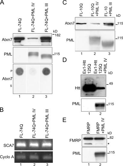
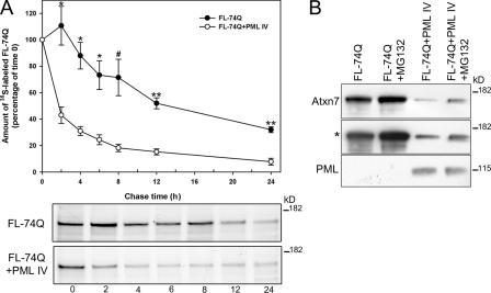
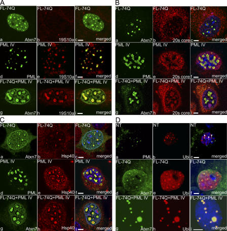
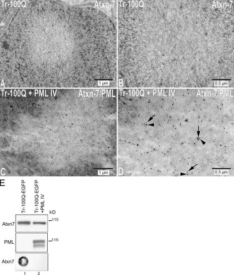
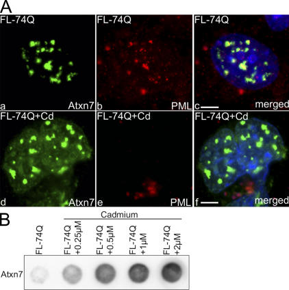
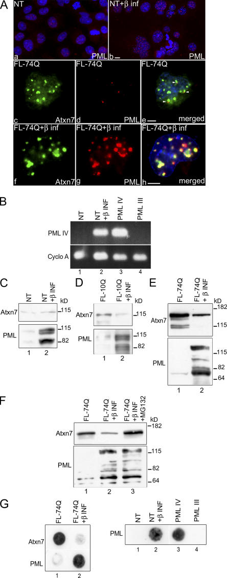
The pathogenesis of spinocerebellar ataxia type 7 and other neurodegenerative polyglutamine (polyQ) disorders correlates with the aberrant accumulation of toxic polyQ-expanded proteins in the nucleus. Promyelocytic leukemia protein (PML) nuclear bodies are often present in polyQ aggregates, but their relation to pathogenesis is unclear. We show that expression of PML isoform IV leads to the formation of distinct nuclear bodies enriched in components of the ubiquitin-proteasome system. These bodies recruit soluble mutant ataxin-7 and promote its degradation by proteasome-dependent proteolysis, thus preventing the aggregate formation. Inversely, disruption of the endogenous nuclear bodies with cadmium increases the nuclear accumulation and aggregation of mutant ataxin-7, demonstrating their role in ataxin-7 turnover. Interestingly, beta-interferon treatment, which induces the expression of endogenous PML IV, prevents the accumulation of transiently expressed mutant ataxin-7 without affecting the level of the endogenous wild-type protein. Therefore, clastosomes represent a potential therapeutic target for preventing polyQ disorders.


Figures

Figure 1.
Figure 1.
ATXN7 aggregates colocalize with PML bodies in SCA7 transgenic mice. Immunofluorescence analysis of cerebellar Purkinje cells (a) and retinal ganglion neurons (b–d) using antibodies against ATXN7 (affinity-purified polyclonal antibody 1261) and PML (chicken anti-PML antibody). Large ATXN7 aggregates sequestered PML bodies (a and b). In ganglion neurons, PML bodies were also surrounded by (b, yellow arrows), colocalized with (c, arrowheads), or juxtaposed to (d, white arrows) small ATXN7 aggregates. The same ganglion neuron on two different focal plans, shown in panels c and d, displayed distinct colocalization between PML bodies and ATXN7 aggregates. Bars, 5 μm.
Figure 2.
Figure 2.
PML isoform IV relocalizes both endogenous and exogenous wild-type and mutant ATXN7. (A) In untransfected cortical neurons (NT), endogenous PML (a) did not colocalize with endogenous ATXN7 (b); note that endogenous nuclear bodies, which can be very small, are not detected in all neurons. Overexpressed PML IV relocalized both endogenous ATXN7 (d–f) and overexpressed wild-type ATXN7 (FL-10Q; g–i) to PML IV bodies. (B) In COS-7 cells, full-length mutant ATXN7 (FL-74Q) partially colocalized with endogenous PML bodies (a–c) and did not colocalize with PML III (d–f) but colocalized with PML IV in large round PML bodies (g–i), some with a ring-like pattern (h, arrowheads). In cortical neurons, FL-74Q and PML IV were also colocalized (j–l). Antibodies used were anti–ATXN7 1C1 and polyclonal anti-PML. Confocal images are shown. Bars, 5 μm.
Figure 3.
Figure 3.
PML IV rapidly recruits soluble mutant ATXN7 from nucleoplasm to PML IV bodies. Time-lapse imaging of living HeLa Kyoto cells expressing EGFP-FL-100Q and RFP-PML IV. 6 h after transfection, cells expressing low levels of EGFP-FL-100Q were monitored every 10 min for a period of 16 h. A series of representative images taken during a 6-h, 40-min time period is shown. From the moment PML IV bodies (red) formed (2 h, 10 min), EGFP-FL-100Q (green) relocalized from the nucleoplasm to these tiny red dots. At 4 h, 20 min, small PML IV bodies coalesced into larger bodies and recruited more EGFP-FL-100Q. At 6 h, 40 min, fusion of ATXN7-containing PML IV bodies was more pronounced, preventing ATXN7 accumulation in the nucleoplasm. Bar, 10 μm.
Figure 4.
Figure 4.
ATXN7 interacts with PML IV. (A and B) Mutant (FL-74Q) or wild-type (FL-10Q) ATXN7 and PML IV were coexpressed in COS-7 cells and immunoprecipitated (IP) or coimmunoprecipitated (Co-IP). Antibodies used were as follows: 1C1 monoclonal anti-ATXN7, PML monoclonal (PMLm), PML polyclonal (PMLp), anti-Xpress (monoclonal negative control), and anti-Myc (polyclonal negative control). (A) Immunoprecipitation of PML IV (lanes 2 and 3) and coimmunoprecipitation of PML IV with ATXN7 (lane 6). Coimmunoprecipitation was not seen with control antibodies (lanes 4, 5, and 7). (B) Immunoprecipitation of mutant ATXN7 (FL-74Q; lane 2) and coimmunoprecipitation with PML (lanes 4 and 5). Coimmunoprecipitation was not seen with control antibodies (lanes 3, 6, and 7). (C) Immunoprecipitation of FL-10Q (lane 2) and coimmunoprecipitation with PML IV (lane 3). Coimmunoprecipitation was not seen with the control antibody (lane 4). The lanes shown in each panel were on the same blots; cuts were made to eliminate lanes irrelevant to the demonstration.
Figure 5.
Figure 5.
ATXN7 and Htt-exon1 are efficiently degraded in the presence of PML IV. (A) Western blot and filter assay of FL-74Q alone or coexpressed with PML IV or III in COS-7 cells. (top) Western blot (30 μg protein, anti–ATXN7 1C1, and anti–PML polyclonal). ATXN7 levels strongly decreased in the presence of PML IV (lane 2) but slightly increased with PML III (lane 3). (bottom) Pellet (P) or supernatant (S) fractions of cell extracts (30 μg protein) were filtered, and SDS-insoluble ATXN7 was revealed by anti–ATXN7 1C1. Coexpression of PML IV led to the disappearance of SDS-insoluble aggregates of mutant ATXN7 (lane 2), whereas PML III slightly increased ATXN7 (lane 3 compared with lane 1). (B) RT-PCR. mRNAs were extracted from cells expressing FL-74Q alone (lane 1) or combined with PML IV (lane 2) or III (lane 3). Primers specific to the SCA7 cDNA and the cyclophilin A (Cyclo A) reference cDNA were used for PCR. Coexpression of PML IV or III had no effect on mutant ATXN7 mRNA. (C) Western blot of cells expressing FL-10Q alone (lane 1) or coexpressed with PML IV (lane 2) or III (lane 3). PML IV decreased and PML III slightly increased ATXN7 levels (same antibodies as in A). (D) Western blot of mutant Htt-exon1 expressed in HeLa cells; Htt-exon1 (Ex1) with 125Q alone (lane 1) or with PML IV (lane 2). Levels of Htt-exon1 (detected with antibody 1C2) were strongly decreased when PML IV was coexpressed. (E) Western blot of FMRP expressed in COS-7 cells alone (lane 1) or with PML IV (lane 2). PML IV did not modify the amount of overexpressed FMRP (detected with antibody 1C3). The asterisk indicates unknown protein degraded in the presence of PML IV. In C, D, and E, the anti-PML antibody was the same as in A.
Figure 6.
Figure 6.
PML IV promotes the degradation of mutant ATXN7 by proteasomes. (A) Pulse-chase analysis of the turnover of FL-74Q expressed alone or with PML IV in COS-7 cells. Quantification of immunoprecipitated ATXN7 from four independent experiments are expressed as means ± SEM. *, P ≤ 0.002; **, P ≤ 0.001 (paired t test). #, P ≤ 0.03 (Mann-Whitney test). (B) Inhibition of the proteasome by MG132 (5 μM) treatment in COS-7 cells significantly increases the amount of FL-74Q expressed alone (compare first and second lanes) or with PML IV (compare third and fourth lanes). The asterisk indicates longer exposure of the same blot. (bottom) PML IV levels.
Figure 7.
Figure 7.
PML IV bodies recruit UPS components. (A) S10a, a subunit of the 19S regulatory complex of the proteasome, was not found in focal aggregates formed by mutant ATXN7 (a–c) but was highly colocalized with PML IV bodies when only PML IV was expressed (d–f). When coexpressed with PML IV, mutant ATXN7 colocalized perfectly with the S10a subunit in PML IV bodies (g–i). (B) The 20S catalytic core of the proteasome faintly colocalized with some ATXN7 aggregates (a–c) but strongly colocalized with PML IV expressed alone (d–f) and with ATXN7 present in PML IV bodies (g–i) when both proteins are coexpressed. (C) The Hsp40 chaperone was found in some ATXN7 aggregates (a–c) but to a much greater extent in PML IV bodies (d–f). Hsp40 also strongly colocalized with ATXN7 when it is relocalized in PML IV bodies (g–i). Confocal images are shown. (D) Polyubiquitinylated proteins (labeled with the FK2 antibody) were enriched in PML IV bodies containing ATXN7. Untransfected cells (NT; a–c) contain a subset of endogenous PML bodies (c, arrowheads) that colocalized with polyubiquitinylated proteins. Aggregates of mutant ATXN7 expressed alone (d–f) colocalized faintly with polyubiquitinylated proteins but strongly colocalized with polyubiquitinylated proteins when recruited in PML IV bodies (g–i). Conventional fluorescence was used in D. Bars, 5 μm.
Figure 8.
Figure 8.
PML IV prevents the formation of fibrillar structures of mutant truncated ATXN7. (A–D) Immunoelectron microscopic images of COS-7 cells expressing Tr-100Q-EGFP alone (A and B) or in combination with PML IV (C and D). The following primary antibodies were used: anti–ATXN7 1C1 (mouse) and anti-PML (rabbit). The following secondary antibodies were used: anti-mouse or anti-rabbit immunoglobulins coupled to 10- or 15-nm gold particles, respectively. ATXN7 (10-nm particles) is seen inside an intranuclear inclusion (A) surrounded by long fibers (higher magnification in B). (C and D) When both PML IV and mutant ATXN7 are expressed, colocalization is observed between ATXN7 (10-nm particles, arrows) and PML (15-nm particles, arrowheads). In double-transfected cells, no fibers can be detected. Nuclear membrane is visible in A (top). Bars: (A and C) 1 μm; (B and D) 0.5 μm. (E) Western blot and filter assay of extracts of cells expressing Tr-100Q-EGFP expressed alone or with PML IV. Coexpression of PML IV led to a modest decrease in the level of soluble Tr-100Q (Western blot) but to a strong decrease in the amount of SDS-insoluble Tr-100Q (filter assay).
Figure 9.
Figure 9.
Cadmium disrupts PML bodies and enhances aggregation of mutant ATXN7. (A) In untreated COS-7 cells, mutant ATXN7 colocalizes with endogenous PML bodies (a–c). In cells treated with 2 μM cadmium (d–f), PML bodies are disrupted and no longer visible (e). Consequently, large ATXN7 aggregates cannot contain any PML bodies (f). (B) The increase in ATXN7 aggregation is dose dependent in cells treated with 0.25–2 μM cadmium (filter assay on pellet fraction of cell extracts; 50 μg).
Figure 10.
Figure 10.
β-INF treatment leads to the degradation of mutant ATXN7 and the disappearance of aggregates. (A) β-INF treatment of COS-7 cells, untransfected (a and b) or expressing FL-74Q (c–h). β-INF increases the number and size of PML bodies (red) in untransfected cells (a and b). FL-74Q aggregates colocalized with some endogenous PML bodies (c–e, arrowheads). β-INF treatment increases both the number and size of PML bodies colocalized with mutant ATXN7 (f–h). Conventional fluorescence was used. Bars, 5 μm. (B) RT-PCR with primers specific to PML IV shows that β-INF increases the expression of endogenous PML IV mRNA in nontransfected (NT) cells (compare lanes 1 and 2), the levels of which are compared with overexpressed PML IV (positive control; lane 3) and overexpressed PML III (negative control; lane 4). The reference transcript cyclophilin A (Cyclo A) was amplified in parallel. (C–E) β-INF induces the expression of multiple isoforms of PML. β-INF does not affect the level of endogenous ATXN7 (C) but decreases the levels of transfected wild-type (D) and mutant ATXN7 (E). (F) PML-dependent degradation of ATXN7 induced by β-INF is inhibited by MG132, showing that it is mediated by the proteasome. (G, left) Filter assay shows that ATXN7 aggregates disappear in β-INF–treated cells, but an insoluble PML-containing fraction appears (lane 2). (right) Insoluble PML material was observed either when PML IV was expressed (lane 3) or when NT cells were treated with β-INF (lane 2), but not when PML III was expressed (lane 4).

Similar articles

Cited by

References

    1. Beech, S.J., K.J. Lethbridge, N. Killick, N. McGlincy, and K.N. Leppard. 2005. Isoforms of the promyelocytic leukemia protein differ in their effects on ND10 organization. Exp. Cell Res. 307:109–117. - PubMed
    1. Bischof, O., O. Kirsh, M. Pearson, K. Itahana, P.G. Pelicci, and A. Dejean. 2002. Deconstructing PML-induced premature senescence. EMBO J. 21:3358–3369. - PMC - PubMed
    1. Borden, K.L. 2002. Pondering the promyelocytic leukemia protein (PML) puzzle: possible functions for PML nuclear bodies. Mol. Cell. Biol. 22:5259–5269. - PMC - PubMed
    1. Cancel, G., C. Duyckaerts, M. Holmberg, C. Zander, G. Yvert, A.S. Lebre, M. Ruberg, B. Faucheux, Y. Agid, E. Hirsch, and A. Brice. 2000. Distribution of ataxin-7 in normal human brain and retina. Brain. 123:2519–2530. - PubMed
    1. Chai, Y., S.L. Koppenhafer, S.J. Shoesmith, M.K. Perez, and H.L. Paulson. 1999. Evidence for proteasome involvement in polyglutamine disease: localization to nuclear inclusions in SCA3/MJD and suppression of polyglutamine aggregation in vitro. Hum. Mol. Genet. 8:673–682. - PubMed

Publication types

MeSH terms