Comparative Study
. 2006 Jul 3;174(1):115-25.
doi: 10.1083/jcb.200602146.

A disease- and phosphorylation-related nonmechanical function for keratin 8

Affiliations
Comparative Study

A disease- and phosphorylation-related nonmechanical function for keratin 8

Nam-On Ku et al. J Cell Biol. .

Abstract

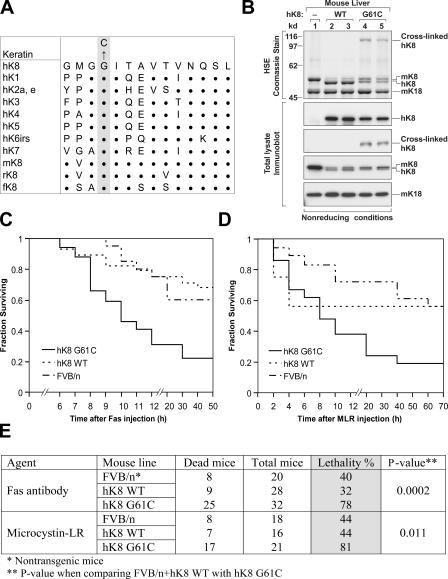
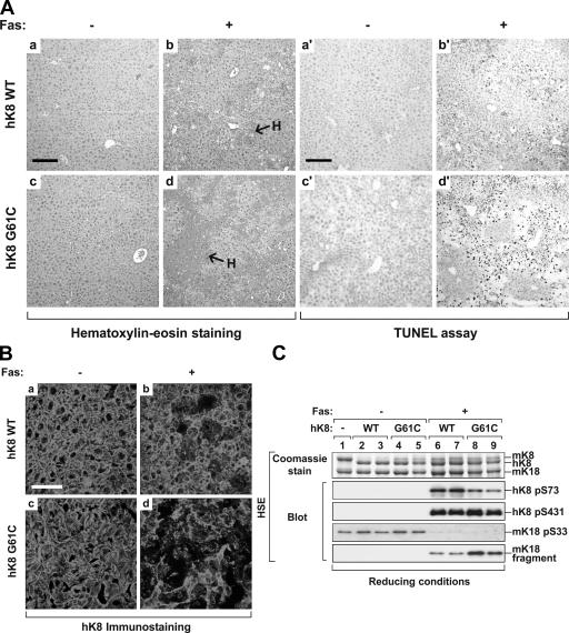
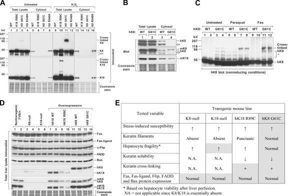
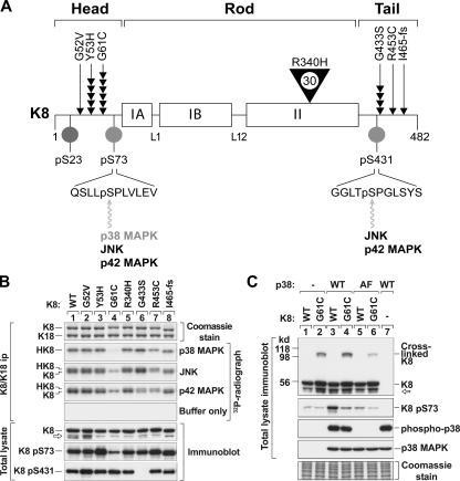
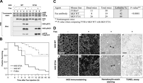
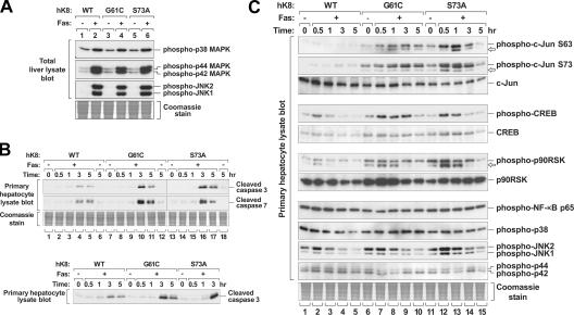
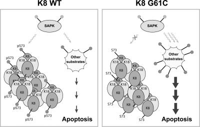
Keratin 8 (K8) variants predispose to human liver injury via poorly understood mechanisms. We generated transgenic mice that overexpress the human disease-associated K8 Gly61-to-Cys (G61C) variant and showed that G61C predisposes to liver injury and apoptosis and dramatically inhibits K8 phosphorylation at serine 73 (S73) via stress-activated kinases. This led us to generate mice that overexpress K8 S73-to-Ala (S73A), which mimicked the susceptibility of K8 G61C mice to injury, thereby providing a molecular link between K8 phosphorylation and disease-associated mutation. Upon apoptotic stimulation, G61C and S73A hepatocytes have persistent and increased nonkeratin proapoptotic substrate phosphorylation by stress-activated kinases, compared with wild-type hepatocytes, in association with an inability to phosphorylate K8 S73. Our findings provide the first direct link between patient-related human keratin variants and liver disease predisposition. The highly abundant cytoskeletal protein K8, and possibly other keratins with the conserved S73-containing phosphoepitope, can protect tissue from injury by serving as a phosphate "sponge" for stress-activated kinases and thereby provide a novel nonmechanical function for intermediate filament proteins.


Figures

Figure 1.
Figure 1.
hK8 G61C mice have increased susceptibility to liver injury. (A) Alignment of the hK8 sequence that is proximal to G61 (shaded residue) with other type II keratins. m, mouse; r, rat; and f, frog. Single letter abbreviations are used for amino acids, and bold dots indicate amino acids that are identical to hK8. (B) Livers were isolated from nontransgenic (lane 1) or transgenic mice that overexpress hK8 WT (lanes 2 and 3) or G61C (lanes 4 and 5). Two independent transgenic lines per genotype were used (i.e., each lane from 2 to 5 represents one mouse per transgenic line). Livers were homogenized, followed by HSE to purify the total keratin pool, or were solubilized in 2% SDS-containing sample buffer to obtain total lysates (Ku et al., 2004). Isolates were separated by SDS-PAGE, and then visualized by Coomassie staining (HSE preparations) or by immunoblotting (total liver homogenates) with Abs to m/hK8, hK8, and mK18. Cross-linked K8 is detected only in livers from K8 G61C mice, and disappears if samples are analyzed under reducing conditions (not depicted). (C and D) The following three mouse genotypes were used: nontransgenic FVB/n, transgenic hK8 WT, or G61C mutant mice (16–32 age- and sex-matched mice per genotype). Each analyzed genotype consisted of nearly equal portions of the two generated independent lines (WT1 and WT2; G61C1 and G61C2). After Fas (C) or MLR (D) injection, mice were assessed hourly for 12 h, and then every 10 h for 3 d. The analyses show survival curves for each administered agent. (E) Summary of the mouse lethality data shown in C and D. No significant difference in mortality was observed when comparing FVB/n with hK8 WT mice, whereas mortality was markedly increased in hK8 G61C mice as compared with the combined FVB/n + hK8 WT mice.
Figure 2.
Figure 2.
K8 G61C accentuates Fas-induced liver injury. Livers were obtained from the indicated transgenic mice 4 h after PBS or Fas Ab administration. (A) Histological analysis (a–d) and TUNEL assay (a′–d′) of transgenic mouse livers that were isolated after PBS (−) or Fas (+) intraperitoneal injection. Note the severe hemorrhage (H) and pyknotic nuclei in hK8 G61C (d and d′) as compared with hK8 WT liver (b and b′) after Fas administration. (B) Immunofluorescence staining of livers similar to those shown in A using an Ab that is specific to hK8. Note the similar staining pattern of hK8 WT and G61C livers under basal conditions (a and c), but the marked hepatocyte drop-off and the collapse of keratin filaments in G61C livers compared with WT mice after Fas injection (b and d). (C) Mouse livers were homogenized, followed by isolation of the total keratin pool by HSE. The extracts were separated by SDS-PAGE, and then visualized by Coomassie staining or blotting with Abs to the indicated epitopes. Each lane represents the analysis of one independent mouse liver. Bars: (A) 160 μm; (B) 40 μm.
Figure 3.
Figure 3.
K8 G61C decreases keratin solubility and promotes keratin cross-linking without altering hepatocyte fragility. (A) BHK cells were transiently cotransfected with the indicated mutant and/or WT keratins (K8 WT + K18 WT; K8 WT + K18 R89C; K18 WT + K8 G61C or R340H). After 3 d, cells were cultured in the presence or absence of 20 mM H2O2 for 1 h, and then divided into two portions. One portion was used to isolate a detergent-free cytosolic soluble fraction and the second to generate a total cell lysate. The isolates were subjected to SDS-PAGE under nonreducing conditions (to visualize cross-linked K8) and blotted with Abs specific to hK8 or hK18. Coomassie staining is also shown to demonstrate equal gel loading. Arrow indicates degraded K8. (B) Liver perfusion was used to isolate hepatocytes from hK8 WT and G61C mice. Total lysates and detergent-free cytosolic fractions were prepared from the isolated hepatocytes, followed by blotting with Abs specific to hK8, m/hK18, and mK18. Open arrows indicate degraded K8. (C) Oxidative stress or apoptosis was induced in mice by intraperitoneal administration of paraquat or Fas Ab, respectively. Total liver lysates were isolated and blotted with anti-hK8 Ab. Asterisk indicates nonspecific bands. Each lane represents the analysis of one independent mouse liver. (D) Total homogenates were prepared from livers of the indicated mouse genotypes (2 mice/genotype), followed by SDS-PAGE and blotting with Abs to the indicated proteins. A duplicate gel was stained with Coomassie blue to verify equal protein loading. Note that the K8 and K18 proteins are absent in both K8- and K18-null livers (lanes 3–6) because of degradation of the partner protein (Baribault et al., 1994; Magin et al., 1998). (E) Liver perfusion of K18-null, K18 R89C, K8 WT, and K8 G61C was performed to assess hepatocyte fragility. K18 R89C was used as a control and hepatocytes from these mice are already known to be fragile (Ku et al., 1995). Hepatocytes from K18-null and hK18 R89C livers had 16–24% viability (n = 3–6 livers/genotype), whereas hK8 WT and G61C hepatocytes had 82–89% viability (n = 4–6 livers/genotype). A summary of the phenotypes of transgenic mice that lack keratin expression (K8- and K18-null) or that express mutant K8 or K18 is shown. Shaded parameters are those examined in this study, whereas unshaded parameters represent summaries of previous studies (Marceau et al., 2001; Omary et al., 2002; Zatloukal et al., 2004).
Figure 4.
Figure 4.
K8 G61C mutation disrupts K8 S73 phosphorylation in vitro and in vivo. (A) Schematic of K8 protein and its in vivo phosphorylation sites (Omary et al., 1998), and the distribution and frequency of K8 mutations associated with human liver disease (Ku et al., 2005). The rod domain of all IF proteins is divided into the following subdomains: IA, linker 1 (L1), IB, L12, and II. Positions of K8 phosphorylation (S23/S73/S431) and the relevant kinases are shown. K8 variants are highlighted by arrowheads, with each arrowhead representing an independent patient from the 467 liver explants that were studied; the single large arrowhead for R340H indicates 30 individuals (Ku et al., 2005). I465-fs is a frame-shift mutation at Ile465 that generates a truncated 468–amino acid protein (instead of 482). (B) BHK cells were transiently cotransfected with K18 WT and one of the indicated K8 constructs, followed by immunoprecipitation of K8/K18, then in vitro phosphorylation by p38, JNK, or p42 kinases. Labeled immunoprecipitates (ip) were analyzed by SDS-PAGE and radiography. HK8 and K8 in the radiograph represent K8 S73 and K8 S431 phosphorylation, respectively. Total lysates were also prepared from the transfected cells then blotted with Abs to the indicated epitopes. No band corresponding to K8 S431 phosphorylation was detected in the pS431 immunoblot of K8 G433S (lane 6) because of an alteration of the Ab epitope in the mutant. (C) BHK cells were triple-transfected with K8/K18 WT or K8 G61C/K18 WT, and WT or kinase-inactive p38 (AF mutant; T180A/Y182F). Total lysates were prepared and blotted with Ab to the indicated epitopes. Open arrows in B and C represent degraded K8.
Figure 5.
Figure 5.
hK8 S73A transgenic mice have increased susceptibility to Fas-mediated hepatocyte apoptosis and injury. (A) Livers from nontransgenic (lane 1) and the indicated transgenic lines (lanes 2–9) were isolated from control or Fas-injected (± Fas) mice and analyzed as described in Fig. 2 C. Two independent transgenic lines per genotype (WT1 and WT2; S73A1 and S73A2) were used for the analysis (each of the lanes 2–9 represents one mouse from individual transgenic lines). (B and C) Nontransgenic FVB/n, hK8 WT, or S73A mice (19–30 age- and sex-matched mice per genotype) were injected with Fas and monitored as described in Fig. 1. Most deaths occurred within 12 h after Fas injection. The results are summarized in C. Mortality was significantly increased in hK8 S73A mice as compared with the combined (FVB/n + hK8 WT) mice. (D) Livers from Fas- or PBS-injected mice were analyzed by immunofluorescence staining (a–b′), by hematoxylin-eosin staining (c and c′), or TUNEL assay (d and d′). Hematoxylin-eosin staining and TUNEL assay of S73A livers under basal conditions (not depicted) were similar to those from the WT livers shown in Fig. 2 A (a and a′). H, hemorrhage. Bars: (a–b′) 40 μm (c–d′) 160 μm.
Figure 6.
Figure 6.
Effect of K8 G61C or S73A mutation on SAPK activation and substrate phosphorylation. (A) Liver homogenates were obtained from the indicated transgenic mice (± Fas injection), separated by SDS-PAGE, and visualized by Coomassie staining (shown for equal loading) or by blotting with the indicated Abs. (B) Hepatocytes were isolated by liver perfusion then treated with 0.5 μg/ml Fas Ab for the indicated times. Total cell lysates were then prepared and analyzed as in A. (top) Lanes 1–12 represent samples loaded on the same gel, whereas lanes 13–18 were loaded on a separate gel. (bottom) Representative samples from the top panel that were analyzed on the same gel to confirm the relative changes in caspase cleavage. (C) Total lysates of primary hepatocytes were prepared and analyzed as in B. Arrows likely represent degradation products. Note that phosphorylation of the SAPK substrates, c-Jun, CREB, and p90RSK, is increased and is more sustained in K8 G61C or S73A hepatocytes as compared with K8 WT hepatocytes.
Figure 7.
Figure 7.
K8 mutation shunts keratin phosphorylation by stress-activated kinases. During stress, activated kinases such as p38 MAPK phosphorylate K8 S73 and other proapoptotic substrates. However, K8 G61C mutation interferes with the ability of the highly abundant K8 S73 to serve as a substrate, which renders other substrates available for potential phosphorylation and consequent rapid progression of apoptosis. Filled dots indicate phosphorylated residues. The phosphate sponge model works in a hepatocyte-protective fashion in livers that express WT K8 and provides a function for K8 S73 phosphorylation that becomes unmasked upon K8 G61C mutation. This model predicts that keratins (and possibly other IFs) undergo hyperphosphorylation in part to absorb, in a “bulk” fashion given their abundance, cell kinase activation. Such sponge activity can be beneficial (e.g., K8 S73), or potentially detrimental, in a phosphorylation site-specific and context-dependent fashion. This model provides one (of several potential) nonmechanical functional capability for IFs and their phosphorylation and does not exclude the possibility that some kinase substrates may undergo dephosphorylation (e.g., via phosphatase activation by phosphorylation) and that other phosphorylation-dependent or -independent mechanisms for protection may be involved.

Similar articles

Cited by

References

    1. Baines, C.P., and J.D. Molkentin. 2005. STRESS signaling pathways that modulate cardiac myocyte apoptosis. J. Mol. Cell. Cardiol. 38:47–62. - PubMed
    1. Baribault, H., J. Penner, R.V. Iozzo, and M. Wilson-Heiner. 1994. Colorectal hyperplasia and inflammation in keratin 8-deficient FVB/N mice. Genes Dev. 8:2964–2973. - PubMed
    1. Behrens, A., M. Sibilia, and E.F. Wagner. 1999. Amino-terminal phosphorylation of c-Jun regulates stress-induced apoptosis and cellular proliferation. Nat. Genet. 21:326–329. - PubMed
    1. Casanova, M.L., A. Bravo, A. Ramirez, G. Morreale de Escobar, F. Were, G. Merlino, M. Vidal, and J.L. Jorcano. 1999. Exocrine pancreatic disorders in transgenic mice expressing human keratin 8. J. Clin. Invest. 103:1587–1595. - PMC - PubMed
    1. Caulin, C., C.F. Ware, T.M. Magin, and R.G. Oshima. 2000. Keratin-dependent, epithelial resistance to tumor necrosis factor–induced apoptosis. J. Cell Biol. 149:17–22. - PMC - PubMed

Publication types

MeSH terms