Comparative Study
. 2006 Jul 3;174(1):127-39.
doi: 10.1083/jcb.200511054.

Signaling across the synapse: a role for Wnt and Dishevelled in presynaptic assembly and neurotransmitter release

Affiliations
Comparative Study

Signaling across the synapse: a role for Wnt and Dishevelled in presynaptic assembly and neurotransmitter release

Azlina Ahmad-Annuar et al. J Cell Biol. .

Abstract

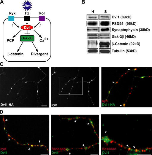
Proper dialogue between presynaptic neurons and their targets is essential for correct synaptic assembly and function. At central synapses, Wnt proteins function as retrograde signals to regulate axon remodeling and the accumulation of presynaptic proteins. Loss of Wnt7a function leads to defects in the localization of presynaptic markers and in the morphology of the presynaptic axons. We show that loss of function of Dishevelled-1 (Dvl1) mimics and enhances the Wnt7a phenotype in the cerebellum. Although active zones appear normal, electrophysiological recordings in cerebellar slices from Wnt7a/Dvl1 double mutant mice reveal a defect in neurotransmitter release at mossy fiber-granule cell synapses. Deficiency in Dvl1 decreases, whereas exposure to Wnt increases, synaptic vesicle recycling in mossy fibers. Dvl increases the number of Bassoon clusters, and like other components of the Wnt pathway, it localizes to synaptic sites. These findings demonstrate that Wnts signal across the synapse on Dvl-expressing presynaptic terminals to regulate synaptic assembly and suggest a potential novel function for Wnts in neurotransmitter release.


Figures

Figure 1.
Figure 1.
Dishevelled colocalizes with presynaptic markers. (A) Diagram illustrating the central role of Dvl in Wnt signaling pathways. PCP, planar cell polarity. Fz, Ryk, and Ror are receptors for Wnts. (B) Analyses of synaptosomes from adult mice brains show the presence of endogenous Dvl at synapses, as well as β-catenin and Gsk-3β. Synaptic proteins such as PSD95 and synaptophysin were used as specific markers for this preparation. H, total brain homogenate; S, total synaptosomal homogenate. (C) Dvl1-HA exhibits a punctate distribution along the axon of hippocampal neurons (9 DIV) as observed with endogenous synapsin I (arrowheads). Merged image shows that many of the Dvl1-HA puncta colocalize with endogenous synapsin I. (D) Staining for endogenous Dvl1 shows that Dvl colocalizes with the presynaptic marker synapsin I and the cytomatrix protein Bassoon (arrowheads). Bar, 10 μm.
Figure 2.
Figure 2.
Wnt signaling through Dvl1 induces synaptic vesicle clustering. (A) MFs exposed to Wnt7b have larger and numerous VAMP2 clusters than control MFs (arrowheads). Addition of the Wnt inhibitor Sfrp-1 to Wnt7b decreases the number of VAMP2 clusters to control levels. (B) 10 DIV hippocampal neurons treated with Wnt7b have more Bassoon puncta than control or Sfrp1 cultures (arrowheads). (C and D) Quantification shows that Wnt7b induces a significant increase in the number of VAMP2 and Bassoon clusters per 100-μm axon length, and that Sfrp-1 blocks this effect to almost control levels. (E) Western blots reveal that Wnt7b does not change the levels of VAMP2, synapsin I, Munc18, syntaxin, or β-catenin proteins in MFs compared with control CM (CCM). (F) Hippocampal neurons were transfected with Dvl1-HA or EGFP before plating and cultured for 9 DIV. Neurons expressing control EGFP exhibit few and faint synapsin I clusters. In contrast, neurons expressing Dvl1-HA have larger and more numerous synapsin I clusters compared with control EGFP (arrowheads). Images at high magnification (bottom) show more synapsin l puncta (arrowheads) in Dvl-HA–expressing neurons than in control EGFP-transfected neurons. (G) Neuron expressing Dvl-HA contains more and larger Bassoon clusters when compared with control EGFP-expressing neurons. (H–I) Quantification shows that Dvl induces a significant increase in the number of synapsin I and Bassoon clusters. Error bars show the SEM. Bars: (A and F) 5 μm; (B) 4 μm; (E) 10 μm; (G) 3 μm.
Figure 3.
Figure 3.
Dvl1 is required presynaptically to mediate Wnt7b signaling. (A) MFs from Dvl1 mutant mice have fewer and smaller VAMP2 clusters than MFs from wild-type mice after 72 h in culture (arrowheads). (B) Quantification shows that Dvl1 mutant MFs exhibit a statistically significant decrease in the number of VAMP2 clusters compared with wild-type MFs (arrowheads). (C) Wild-type and Dvl1 mutant MFs were treated with control CM (CCM) and Wnt7b CM for 16 h. Wnt7b only partially rescues the number of VAMP2 puncta in Dvl1 mutant MFs. (D) Quantification shows that Wnt7b induces a 40% increase in the number of VAMP2 puncta in wild-type MFs, but only a 20% increase in Dvl1 mutant MFs. Graphs show the number of synaptic vesicle puncta per 100 μm neurite length ± the SEM. Error bars show the SEM. Asterisks indicate *, P < 0.05; **, P < 0.01. Bars, 5 μm.
Figure 4.
Figure 4.
Recycling activity of synaptic vesicles is mediated by Wnt signaling. Synaptic vesicle recycling in MFs are visualized by the uptake of FM1-43 or intralumenal synaptotagmin (syt) antibody and counterstained with SV2. (A and C) Wnt7b increases the number of anti-synaptotagmin and FM1-43 recycling sites (arrowheads) compared with control MF axons, and the presence of Sfrp-1 blocks this effect. For synaptotagmin uptake, merged figures show colocalization of puncta labeled with anti-synaptotagmin with the synaptic vesicle protein SV2 (arrowheads). (B and D) Quantification shows that Wnt7b increases the number of recycling sites of anti-synaptotagmin antibody and FM1-43 uptake per 100-μm neurite length whereas Sfrp-1 blocks the activity of Wnt7b. (E) Western blot reveals that Wnt7b does not affect the level of synaptotagmin protein in MFs compared with control CM (CCM). (F) Dvl1 / mutant MFs have fewer synaptic vesicle recycling sites when compared with wild-type MFs (arrowheads). (G) Quantification of the number of recycling sites per 100-μm neurite length indicates a 37% decrease in MFs from Dvl1 / mutants when compared with wild-type. Error bars show the SEM. *, P <0.05; **, P < 0.01. Bars: (A and C) 10 μm; (F) 5 μm.
Figure 5.
Figure 5.
Wnt7a//Dvl1/ double mutant mice have smaller areas of synapsin l staining at glomerular rosettes at P10 and P15. (A) At P10, Wnt7a and Dvl1 single mutant mice exhibit a significant decrease in the number of stained glomerular rosettes when compared with wild type. This effect is significantly enhanced in Wnt7a /; Dvl1 / double mutant mice as the areas are less stained and smaller. (B) Size distribution of glomerular rosettes stained with synapsin I at P10 shows a significant increase in the proportion of glomerular rosettes with a size <40 μm2 in the double mutants (ANOVA; P < 0.01) when compared with wild type, and a concomitant decrease in stained rosettes with a size >80 μm2. Single mutants also exhibit a decrease in the smaller stained rosettes with a concomitant decrease in the large ones. (C) At P15, no significant differences are found between wild-type and single Wnt7a and Dvl1 mutants. However, the double mutant still exhibits a defect. Arrows, big rosettes; arrowheads, small rosettes. (D) Size distribution of glomerular rosettes stained with synapsin I at P15 shows no differences between single mutants and wild type. However, Wnt7a /;Dvl1 / double mutants still exhibit a defect with a significantly larger proportion of stained rosettes with a size <40 μm2. Values are mean ± SEM. *, P < 0.05; **, P < 0.01; ***, P < 0.001. Bar, 20 μm.
Figure 6.
Figure 6.
Cerebellar glomerular rosettes are less complex in Wnt7a; Dvl1 double mutants. Electron micrographs of P15 wild-type (A), Wnt7a single mutant (B), Dvl1 single mutant (C), and Wnt7a //Dvl1 / double mutant (D) and their respective outlines (A'–D') show the morphology of glomerular rosettes. Single mutants have complex rosettes similar to wild type (compare A, B, and C). However, the morphology of glomerular rosettes is less irregular in the Wnt7a //Dvl1 / double mutant (D). Quantification reveals a significant decrease in the complexity (measured by perimeter2/area) of MF terminals from double mutant animals when compared with wild-type (P < 0.01). In A'–D', asterisks indicate interdigitations in the MF terminal. Values in each image represent the complexity of the terminal. Three independently bred wild-type, Wnt7a /, and Dvl1 / mice and five Wnt7a //Dvl1 / double mutant animals were analyzed. 70–180 rosettes were measured per genotype. GC, GC dendrite. Bar, 0.5 μm.
Figure 7.
Figure 7.
The frequency of mEPSCs is reduced in Wnt7a; Dvl1 double mutants. Spontaneous EPSCs made from GCs of P15 mice. A representative whole-cell voltage clamp recording made from a GC in a cerebellar slice prepared from wild-type (A) and Wnt7a /;Dvl1 / (D) double mutant animals. In each image, detected mEPSCs (large transient inward deflections) are marked with gray circles on the continuous current record. (B and E) Superimposed spontaneous EPSCs from wild-type (B) and double mutant (E) animals. (G) Histogram shows the frequency of spontaneous EPSC from wild-type and Wnt7a /; Dvl1 / double mutant animals. Fewer spontaneous events occur in the Wnt7a /; Dvl1 / double mutant (n = 8) when compared with wild type (n = 6; unpaired t test). (C and F) Representative amplitude distributions from wild-type (C) and Wnt7a /; Dvl1 / double mutant (F) mice. (H) Histogram shows no significant differences in the amplitude of mEPSC between wild-type (−10.8 ± 2.2 pA) and Wnt7a /; Dvl1 / double mutant (−8.7 ± 0.9 pA) animals. Error bars indicate the SEM (unpaired t test).

References

    1. Cadigan, K.M., and Y.I. Liu. 2006. Wnt signaling: complexity at the surface. J. Cell Sci. 119:395–402. - PubMed
    1. Cathala, L., S. Brickley, S. Cull-Candy, and M. Farrant. 2003. Maturation of EPSCs and intrinsic membrane properties enhances precision at a cerebellar synapse. J. Neurosci. 23:6074–6085. - PMC - PubMed
    1. Christopherson, K.S., E.M. Ullian, C.C. Stokes, C.E. Mullowney, J.W. Hell, A. Agah, J. Lawler, D.F. Mosher, P. Bornstein, and B.A. Barres. 2005. Thrombospondins are astrocyte-secreted proteins that promote CNS synaptogenesis. Cell. 120:421–433. - PubMed
    1. Ciani, L., and P.C. Salinas. 2005. WNTs in the vertebrate nervous system: from patterning to neuronal connectivity. Nat. Rev. Neurosci. 6:351–362. - PubMed
    1. Ciani, L., O. Krylova, M. Smalley, T. Dale, and P.C. Salinas. 2003. A divergent canonical WNT signalling pathway regulates microtubule dynamics: Dishevelled signals locally to stabilize microtubules. J. Cell Biol. 164:243–253. - PMC - PubMed

Publication types

MeSH terms