Comparative Study
. 2006 Jul 15;177(2):925-33.
doi: 10.4049/jimmunol.177.2.925.

Antigen-experienced T cells limit the priming of naive T cells during infection with Leishmania major

Affiliations
Comparative Study

Antigen-experienced T cells limit the priming of naive T cells during infection with Leishmania major

Peter M Gray et al. J Immunol. .

Abstract

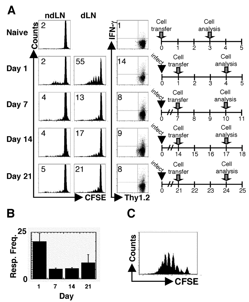
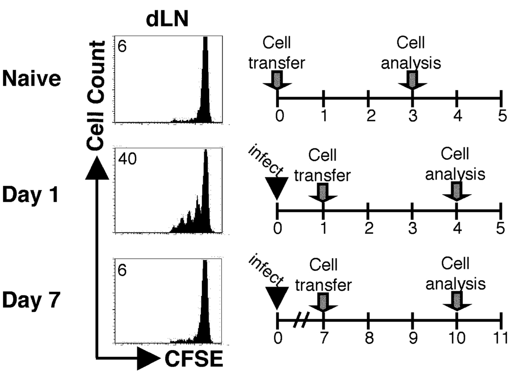
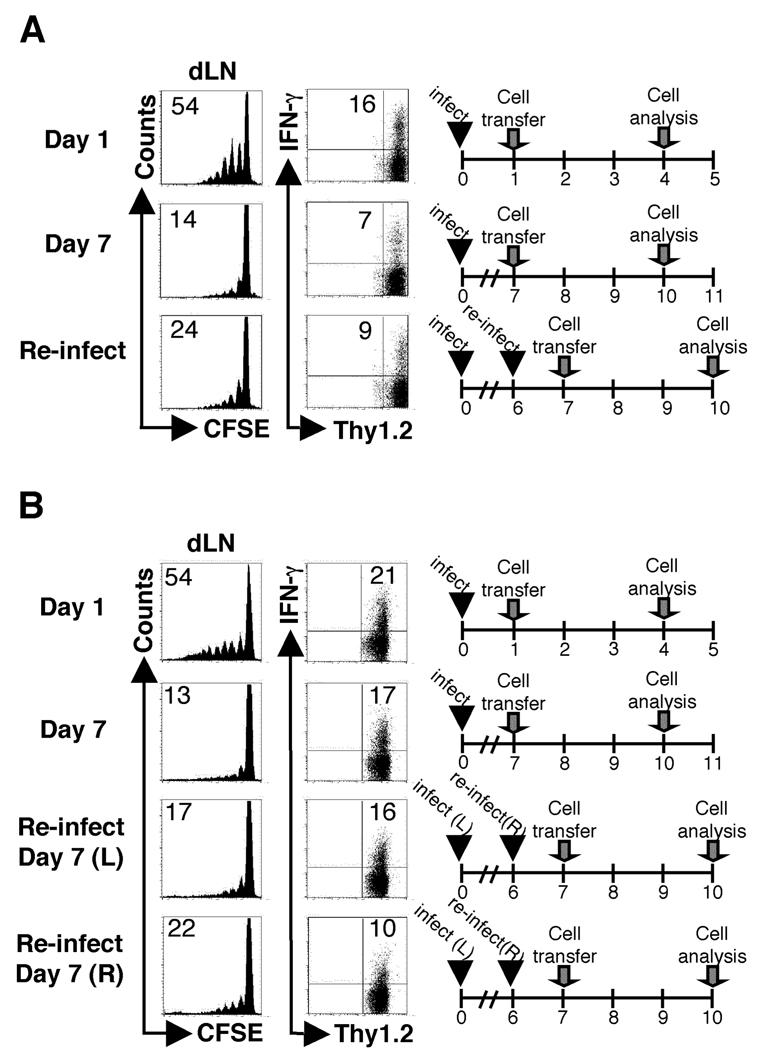
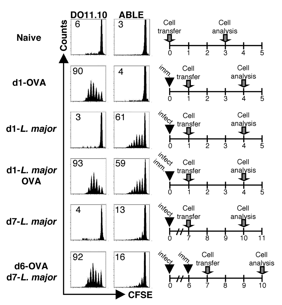
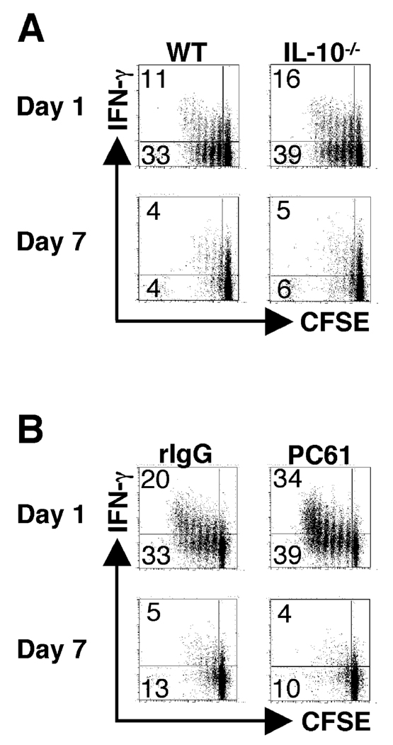
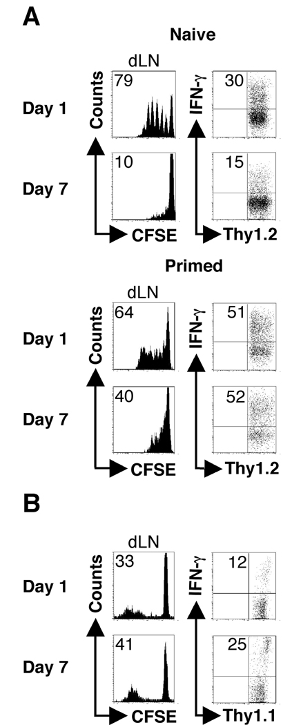
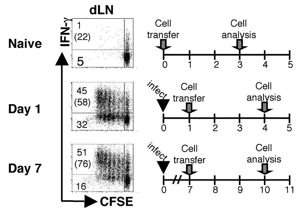
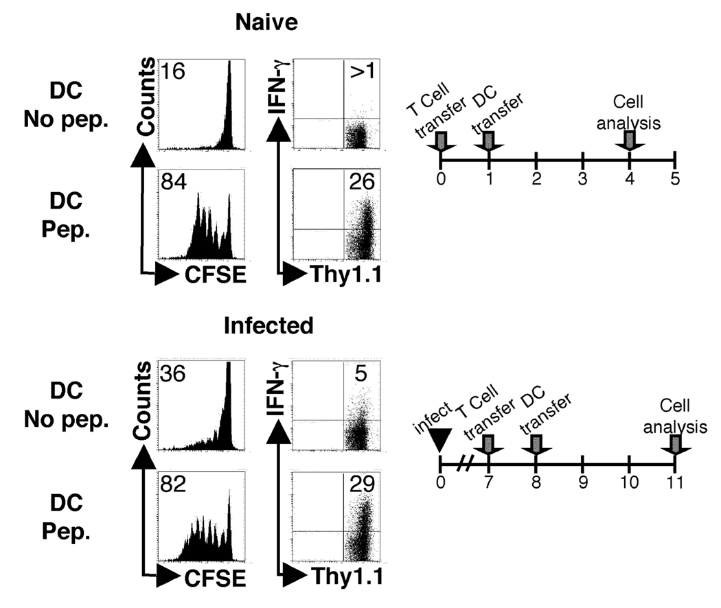
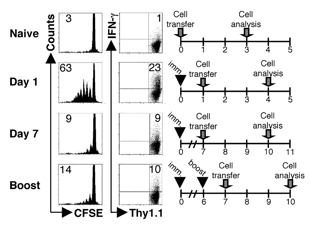
One mechanism to control immune responses following infection is to rapidly down-regulate Ag presentation, which has been observed in acute viral and bacterial infections. In this study, we describe experiments designed to address whether Ag presentation is decreased after an initial response to Leishmania major. Naive alphabeta-Leishmania-specific (ABLE) TCR transgenic T cells were adoptively transferred into mice at various times after L. major infection to determine the duration of presentation of parasite-derived Ags. ABLE T cells responded vigorously at the initiation of infection, but the ability to prime these cells quickly diminished, independent of IL-10, regulatory T cells, or Ag load. However, Ag-experienced clonal and polyclonal T cell populations could respond, indicating that the diminution in naive ABLE cell responses was not due to lack of Ag presentation. Because naive T cell priming could be restored by removal of the endogenous T cell population, or adoptive transfer of Ag-pulsed dendritic cells, it appears that T cells that have previously encountered Ag during infection compete with naive Ag-specific T cells. These results suggest that during L. major infection Ag-experienced T cells, rather than naive T cells, may be primarily responsible for sustaining the immune response.


Figures

Figure 1
Figure 1
The ability to activate naïve L. major-specific T cells rapidly diminishes following infection. BALB/c recipient mice (Thy1.1+) were infected with 5×10⁶ stationary phase L. major promastigotes and CFSE-labeled ABLE transgenic T cells (Thy1.2+) were transferred at the indicated times following infection. Three days following transfer, cells were harvested and analyzed by flow cytometry. (A) Histogram plots were gated on donor cells (Thy1.2+). The number in the corner represents the percentage of donor cells that were CFSEdim in the non-draining or dLNs. The numbers in the corners of the dot plots indicated the percentage of donor cells that acquired the ability to produce IFN-γ in the dLN of the recipient animals. (B) The percentage of donor cells that responded by proliferating in the dLN at each time point was calculated (see Materials and Methods). (C) Whole dLNs from mice that received ABLE cells seven days after infection and harvested 3 days later were isolated and restimulated in vitro with 1µg of LACK peptide for 3 days. Donor cells were gated on by Thy1.2 expression. The data are representative of three or more experiments.
Figure 2
Figure 2
The ability to activate naïve T cells specific for an alternative antigen rapidly diminishes following infection. BALB/c recipient mice (Thy1.2+) were infected with transgenic Leishmania promastigotes expressing ovalbumin (Leishmania-OVA) and CFSE-labeled DO11.10 transgenic T cells were transferred at the indicated times following infection. Three days following transfer, cells were harvested and analyzed by flow cytometry. Histogram plots were gated on donor cells (KJ126+CD4+). The number in the corner represents the percentage of donor cells that were CFSEdim in the non-draining or dLNs.
Figure 3
Figure 3
Subsequent exposure to live parasites does not rescue the inability to activate naïve cells at d7. BALB/c recipient mice (Thy1.1+) were infected as described in Fig. 1. At d6 post primary infection one group of mice was re-infected in the same (A) or contralateral (B) footpad with L. major, control mice were given PBS in place of parasites at d6. CFSE-labeled ABLE transgenic T cells (Thy1.2+) were adoptively transferred following infection as indicated. Cells were harvested and analyzed as in Fig. 1 with the exception that the primary (L) and secondary (R) dLNs were analyzed from mice challenged in the contralateral footpad. Donor cells were gated on by Thy1.2 expression. The data are representative of three experiments.
Figure 4
Figure 4
Antigen presentation to an unrelated antigen is intact at seven days following L. major infection. BALB/c recipient mice (Thy1.2+) were infected with L. major. Six days following infection one group of mice was injected with 25ug of OVA protein in the same footpad (d6-OVA/d7-Leish). As controls, mice were given protein alone (d1-OVA), infected with L. major for one or seven days (d1-Leish and d7-Leish, respectively), or infected and immunized at the same time (d1-Leish/OVA). CFSE-labeled OVA-specific (DO11.10) transgenic T and ABLE Thy1.1+ cells were adoptively transferred into the recipient mice at the indicated time points. The dLNs were harvested and analyzed by flow cytometry three days following transfer. Histogram plots were gated on KJ1-26+CD4+ (DO11.10) or Thy1.1+ (ABLE) donor cells in the dLNs of recipient mice. The data are representative of three experiments.
Figure 5
Figure 5
The suboptimal ability to activate naïve T cells at seven days following L. major infection is not dependent upon immunosuppression. (A) BALB/c IL-10−/− recipient mice (Thy1.2+) were infected with L. major and CFSE-labeled ABLE transgenic T cells (Thy1.1+) were transferred at one or seven days following infection. As controls, experiments were also performed with wild type recipient mice as described in Fig. 1. Dot plots are gated on donor cells in the dLNs of recipient mice (Thy1.1+). The numbers in the corners of the dot plots represent the percentage of events in either of the CFSEdim quadrants, and the numbers in parentheses represent the percentage of cells that produce IFN-γ that are CFSEdim. (B) BALB/c recipient mice (Thy1.2) were depleted of Tregs by administration of 1mg of anti-CD25 (PC61) antibody intraperitoneally 7 days prior to infection, and control mice were treated with 1mg of rat IgG. Following infection, CFSE-labeled ABLE transgenic T cells (Thy1.1+) cells were transferred, harvested, and analyzed as described in Fig. 1. Dot plots were gated on donor cells in the dLNs of recipient mice (Thy1.1+).
Figure 6
Figure 6
Previously activated ABLE transgenic T cells and immune polyclonal cells are less susceptible to the suboptimal activation observed seven days following infection. (A) BALB/c recipient mice (Thy1.1+) were infected as described in Fig. 1. CFSE-labeled ABLE transgenic naïve or previously activated (primed)(Thy1.2+)(see Materials and Methods) were adoptively transferred into recipient mice at d1 or d7 following infection. The dLNs were harvested and analyzed by flow cytometry three days following transfer. Histogram plots were gated on donor cells (Thy1.2+). The number in the corner represents the percentage of donor cells that were CFSEdim in the dLNs. The numbers in the corners of the dot plots indicated the percentage of donor cells that acquired the ability to produce IFN-γ in the dLN of the recipient animals. (B) Naïve C57BL/6 mice (Thy1.2+) were infected as described in Fig. 1. Immune cells from C57BL/6 mice (Thy1.1+) that had healed infection (>12weeks following infection) were transferred at the indicated times following infection. Seven days following transfer cells were harvested and analyzed by flow cytometry. Donor cells were gated on by Thy1.1 expression. The data are representative of three experiments.
Figure 7
Figure 7
The previously activated antigen-specific cells prevent the optimal activation of naïve antigen-reactive cells. DO11.10 RAG2−/− recipient mice (Thy1.2+) were infected as described in Fig. 1. CFSE-labeled ABLE transgenic cells (Thy1.1+) were adoptively transferred into recipient mice at d1 or d7 following infection. The dLNs of recipient mice were harvested and analyzed by flow cytometry three days following transfer. Dot plots were gated on donor cells in the dLNs of recipient mice (Thy1.1+). The numbers in the corners represent the percentage of events in either of the CFSEdim quadrants, and the numbers in parentheses represent the percentage of cells that produce IFN-γ that are CFSEdim. The data are representative of three experiments.
Figure 8
Figure 8
Naïve and antigen-experienced T cells compete for access to antigen presenting cells. Naïve BALB/c mice (Thy1.2+) were either infected with 5e6 stationary phase promastigotes or left uninfected. Naïve CFSE-labeled ABLE transgenic T cells (Thy1.1+) were transferred into naïve or d7 infected mice. One day after transfer LPS matured BMDDC pulsed with 5ug of LACK peptide or left unpulsed were adoptively transferred directly into the recipient via the footpad. Three days following transfer of BMDDC, cells were harvested and analyzed by flow cytometry. Histogram plots were gated on donor cells (Thy1.1+). The number in the corner represents the percentage of donor cells that were CFSEdim in the dLNs. Non-draining lymph nodes were also harvested and analyzed for proliferation (insets). The numbers in the corners of the dot plots indicated the percentage of donor cells that acquired the ability to produce IFN-gamma in the dLN of the recipient animals. The data are representative of two experiments.
Figure 9
Figure 9
Secondary immunizations recruit few naïve T cells into the immune response. BALB/c recipient mice (Thy1.2+) were immunized in the hind footpad with 50µg of soluble Leishmania antigen (SLA) and 50µg of CpG DNA. In addition, a third group of mice was boosted with a second administration of SLA and CpG DNA in the same footpad at d6 following primary immunization (boost). CFSE-labeled ABLE transgenic T cells (Thy1.1+) were transferred at either d1 or d7 following the primary immunization. As a negative control, ABLE transgenic T cells were transferred into naïve BALB/c mice. The data are representative of two independent experiments.

Similar articles

Cited by

References

    1. Sacks D, Noben-Trauth N. The immunology of susceptibility and resistance to Leishmania major in mice. Nat Rev Immunol. 2002;2:845–858. - PubMed
    1. Zaph C, Uzonna J, Beverley SM, Scott P. Central memory T cells mediate long-term immunity to Leishmania major in the absence of persistent parasites. Nat Med. 2004;10:1104–1110. - PubMed
    1. Wong P, Pamer EG. Feedback regulation of pathogen-specific T cell priming. Immunity. 2003;18:499–511. - PubMed
    1. van Faassen H, Dudani R, Krishnan L, Sad S. Prolonged antigen presentation, APC−, and CD8+ T cell turnover during mycobacterial infection: comparison with Listeria monocytogenes. J Immunol. 2004;172:3491–3500. - PubMed
    1. van Faassen H, Saldanha M, Gilbertson D, Dudani R, Krishnan L, Sad S. Reducing the stimulation of CD8+ T cells during infection with intracellular bacteria promotes differentiation primarily into a central (CD62LhighCD44high) subset. J Immunol. 2005;174:5341–5350. - PubMed

Publication types

MeSH terms

Substances