. 2006 Jul 11;103(28):10817-22.
doi: 10.1073/pnas.0602754103. Epub 2006 Jul 3.

A phosphatidic acid-binding protein of the chloroplast inner envelope membrane involved in lipid trafficking

Affiliations

A phosphatidic acid-binding protein of the chloroplast inner envelope membrane involved in lipid trafficking

Koichiro Awai et al. Proc Natl Acad Sci U S A. .

Abstract

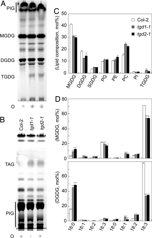
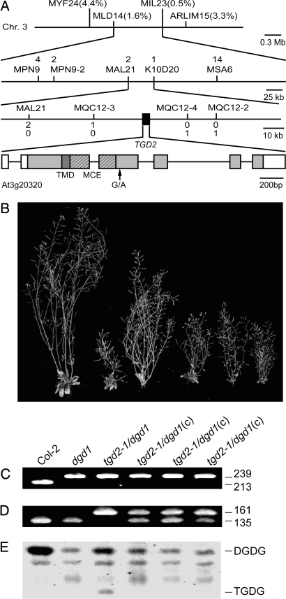
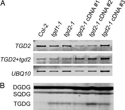
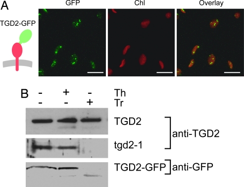
The biogenesis of the photosynthetic thylakoid membranes inside plant chloroplasts requires enzymes at the plastid envelope and the endoplasmic reticulum (ER). Extensive lipid trafficking is required for thylakoid lipid biosynthesis. Here the trigalactosyldiacylglycerol2 (tgd2) mutant of Arabidopsis is described. To the extent tested, tgd2 showed a complex lipid phenotype identical to the previously described tgd1 mutant. The aberrant accumulation of oligogalactolipids and triacylglycerols and the reduction of molecular species of galactolipids derived from the ER are consistent with a disruption of the import of ER-derived lipids into the plastid. The TGD1 protein is a permease-like component of an ABC transporter located in the chloroplast inner envelope membrane. The TGD2 gene encodes a phosphatidic acid-binding protein with a predicted mycobacterial cell entry domain. It is tethered to the inner chloroplast envelope membrane facing the outer envelope membrane. Presumed bacterial orthologs of TGD1 and TGD2 in Gram-negative bacteria are typically organized in transcriptional units, suggesting their involvement in a common biological process. Expression of the tgd2-1 mutant cDNA caused a dominant-negative effect replicating the tgd2 mutant phenotype. This result is interpreted as the interference of the mutant protein with its native protein complex. It is proposed that TGD2 represents the substrate-binding or regulatory component of a phosphatidic acid/lipid transport complex in the chloroplast inner envelope membrane.


Conflict of interest statement

Conflict of interest statement: No conflicts declared.

Figures

Fig. 1.
Fig. 1.
Lipid phenotype of the tgd2-1 mutant compared with the tgd1-1 mutant and the Col-2 wild type. (A) Thin-layer chromatogram of polar lipids. Lipids were visualized by α-naphthol staining. (B) Thin-layer chromatogram of neutral lipids. Lipids were visualized by exposure to iodine vapor. (C) Polar lipid composition (relative mol%) determined by quantification of fatty acid methylesters derived from individual lipids. (D) Fatty acid composition of the two galactolipids MGDG and DGDG. The legend is the same as that for C. Fatty acids are indicated with number of carbons:number of double bonds. DGDG, digalactosyldiacylglycerol; MGDG, monogalactosyldiacylglycerol; O, origin; PC, phosphatidylcholine; PE phosphatidylethanolamine; PG, phosphatidylglycerol; PI, phosphatidylinositol; PIG, pigments; SQDG, sulfoquinovosyldiacylglycerol; TAG, triacylglycerol; TGDG, trigalactosyldiacylglycerol.
Fig. 2.
Fig. 2.
Identification of the TGD2 locus. (A) Map position of the tgd2-1 mutation on chromosome 3 and structure of the TGD2 gene (At3g20320). Markers used for mapping and the respective number of recombinations are indicated. The TGD2 gene is indicated by a black box and expanded on the lowest line. The coding region of At3g20320 is shown as a shaded box. The darker shading indicates the predicted TMD. A region encoding an MCE domain is shown hashed. Introns are indicated by a line. Noncoding regions of the gene deduced from the cDNA are shown as open boxes. (B) Growth of different plants on soil (8 weeks old) with a genotype as indicated below the panel. Mutants were homozygous at all indicated loci. Three plants from independent transformation events expressing the TGD2 cDNA are indicated by “(c).” (C and D) Genotyping at the DGD1 locus (C) and at the TGD2 locus (D). Point mutation-specific dCAPS markers were used, and ethidium bromide-stained DNA diagnostic DNA fragments are shown with their respective lengths in base pairs. (E) Lipid phenotype of the six different plant lines. A section of thin-layer chromatogram stained for glycolipids is shown. DGDG, digalactosyldiacylglycerol; TGDG, trigalactosyldiacylglycerol.
Fig. 3.
Fig. 3.
Expression of the tgd2-1 mutant cDNA in the Col-2 wild type. The untransformed wild type (Col-2) and the untransformed tgd1-1 and tgd2-1 mutants are included for comparison. Three independent transformants are shown. (A) Semiquantitative RT-PCR of mRNA levels derived from the TGD2 wild-type gene (Top), the TGD2 wild-type gene and the tgd2-1 transgene (Middle), and the ubiquitin (UBQ10) control (Bottom). Negative images of ethidium bromide-stained gels are shown. (B) Polar lipid phenotype of the indicated plants. A section of the thin-layer chromatogram stained for glycolipids is shown. DGDG, digalactosyldiacylglycerol; SQDG, sulfoquinovosyldiacylglycerol; TGDG, trigalactosyldiacylglycerol.
Fig. 4.
Fig. 4.
Subcellular localization and topology of TGD2 after transient expression in tobacco leaves. (A) Localization of full-length TGD2 protein fused to GFP (TGD2–GFP). The insertion of the respective protein into the membrane is schematically shown on the left. GFP, green fluorescence specific for GFP; Chl, red fluorescence of chloroplasts; the overlay of the two images is shown on the right. Confocal images are shown. (Scale bars: 10 μm.) (B) Topology of the TGD2 protein. The wild-type TGD2 protein, the tgd2-1 mutant protein, and the GFP fusion were transiently produced in tobacco leaves, and isolated chloroplasts were analyzed. The TGD2 and tgd2-1 proteins were detected by using a TGD2-specific antibody. The GFP fusion was detected by using a GFP-specific antibody. Samples were untreated with protease (−) or treated with thermolysin (+, Th) or with trypsin (+, Tr). Immunoblots are shown.
Fig. 5.
Fig. 5.
Binding of recombinant TGD2 protein to lipids. The TGD2 protein is N-terminally truncated lacking the TMD to exclude lipid binding to this region of the protein. (A and B) Membrane binding assay with commercial phospholipid-containing membrane (A) or plant lipid-containing membrane (B). (C) Liposome binding assay. Liposomes consisted of phosphatidylcholine only (PC, first lane) or PC (60% wt/wt, second through fourth lanes) mixed with different molecular species of PA (40% wt/wt). PA molecular species tested were dioleoyl-PA (18:1), sn1-oleoyl, sn2-palmitoyl PA (18:1/16:0), and dipalmitoyl-PA (16:0). DGDG, prokaryotic digalactosyldiacylglycerol; DGDGe, eukaryotic digalactosyldiacylglycerol; L-PA, lysophosphatidic acid; L-PC, lysophosphatidylcholine; MGDG, prokaryotic monogalactosyldiacylglycerol; MGDGe, eukaryotic monogalactosyldiacylglycerol; PC, phosphatidylcholine; PE, phosphatidylethanolamine; PG, phosphatidylglycerol; PI, phosphatidylinositol; PIP(3), phosphatidylinositol 3-phosphate; PIP(4), phosphatidylinositol 4-phosphate; PIP(5), phosphatidylinositol 5-phosphate; PIP2(3,4), phosphatidylinositol 3,4-bisphosphate; PIP2(3,5), phosphatidylinositol 3,5-bisphosphate; PIP2(4,5), phosphatidylinositol 4,5-bisphosphate; PIP3(3,4,5), phosphatidylinositol 3,4,5-bisphosphate; PS, phosphatidylserine; S1P, sphingosine 1-phosphate; SQDG, sulfoquinovosyldiacylglycerol; TGDG, trigalactosyldiacylglycerol.

Similar articles

Cited by

References

    1. Browse J., Somerville C. Annu. Rev. Plant Physiol. Plant Mol. Biol. 1991;42:467–506.
    1. Roughan P. G., Slack C. R. Annu. Rev. Plant Physiol. 1982;33:97–132.
    1. Browse J., Warwick N., Somerville C. R., Slack C. R. Biochem. J. 1986;235:25–31. - PMC - PubMed
    1. Benning C., Ohta H. J. Biol. Chem. 2005;280:2397–2400. - PubMed
    1. Kunst L., Browse J., Somerville C. Proc. Natl. Acad. Sci. USA. 1988;85:4143–4147. - PMC - PubMed

Publication types

MeSH terms

Associated data