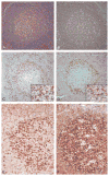
Programmed death-1 (PD-1) is a marker of germinal center-associated T cells and angioimmunoblastic T-cell lymphoma
- PMID: 16819321
- PMCID: PMC3137919
- DOI: 10.1097/01.pas.0000209855.28282.ce
Programmed death-1 (PD-1) is a marker of germinal center-associated T cells and angioimmunoblastic T-cell lymphoma
Abstract
Programmed death-1 (PD-1), a member of the CD28 costimulatory receptor family, is expressed by germinal center-associated T cells in reactive lymphoid tissue. In a study of a wide range of lymphoproliferative disorders, neoplastic T cells in 23 cases of angioimmunoblastic lymphoma were immunoreactive for PD-1, but other subtypes of T cell and B cell non-Hodgkin lymphoma, as well as classic Hodgkin lymphoma, did not express PD-1. The pattern of PD-1 immunostaining of neoplastic cells in angioimmunoblastic lymphoma was similar to that reported for CD10, a recently described marker of neoplastic T cells in angioimmunoblastic lymphoma. Tumor-associated follicular dendritic cells in cases of angioimmunoblastic lymphoma were found to express PD-L1, the PD-1 ligand. In addition, PD-1-positive reactive T cells formed rosettes around neoplastic L&H cells in 14 cases of nodular lymphocyte predominant Hodgkin lymphoma studied. These findings, along with data from previous studies, suggest that angioimmunoblastic lymphoma is a neoplasm of germinal center-associated T cells and that there is an association of germinal center-associated T cells and neoplastic cells in nodular lymphocyte predominant Hodgkin lymphoma. PD-1 is a useful new marker for angioimmunoblastic lymphoma and lends further support to a model of T-cell lymphomagenesis in which specific subtypes of T cells may undergo neoplastic transformation and result in specific, distinct histologic, immunophenotypic, and clinical subtypes of T-cell neoplasia.
Figures


Similar articles
-
Expression of two markers of germinal center T cells (SAP and PD-1) in angioimmunoblastic T-cell lymphoma.Haematologica. 2007 Aug;92(8):1059-66. doi: 10.3324/haematol.10864. Epub 2007 Jul 20. Haematologica. 2007. PMID: 17640856
-
Angioimmunoblastic T-cell lymphoma with hyperplastic germinal centres: a neoplasia with origin in the outer zone of the germinal centre? Clinicopathological and immunohistochemical study of 10 cases with follicular T-cell markers.Mod Pathol. 2009 Jun;22(6):753-61. doi: 10.1038/modpathol.2009.12. Epub 2009 Mar 27. Mod Pathol. 2009. PMID: 19329936
-
Germinal-center T-helper-cell markers PD-1 and CXCL13 are both expressed by neoplastic cells in angioimmunoblastic T-cell lymphoma.Am J Clin Pathol. 2009 Jan;131(1):33-41. doi: 10.1309/AJCP62WRKERPXDRT. Am J Clin Pathol. 2009. PMID: 19095563
-
The life and death of the germinal center.Ann Diagn Pathol. 2020 Feb;44:151421. doi: 10.1016/j.anndiagpath.2019.151421. Epub 2019 Nov 13. Ann Diagn Pathol. 2020. PMID: 31751845 Review.
-
[Research advances of PD-1 in angioimmunoblastic T-cell lymphoma].Zhonghua Bing Li Xue Za Zhi. 2024 Sep 8;53(9):973-976. doi: 10.3760/cma.j.cn112151-20240226-00120. Zhonghua Bing Li Xue Za Zhi. 2024. PMID: 39231759 Review. Chinese.
Cited by
-
Cerebellar EBV-associated diffuse large B cell lymphoma following angioimmunoblastic T cell lymphoma.J Hematop. 2015 Dec;8(4):235-241. doi: 10.1007/s12308-015-0241-8. Epub 2015 Mar 18. J Hematop. 2015. PMID: 27559423 Free PMC article.
-
Catching up with solid tumor oncology: what is the evidence for a prognostic role of programmed cell death-ligand 1/programmed cell death-1 expression in B-cell lymphomas?Haematologica. 2016 Oct;101(10):1144-1158. doi: 10.3324/haematol.2016.145904. Haematologica. 2016. PMID: 27694502 Free PMC article. Review.
-
Recent advances in understanding and managing T-cell lymphoma.F1000Res. 2017 Dec 12;6:2123. doi: 10.12688/f1000research.12573.1. eCollection 2017. F1000Res. 2017. PMID: 29259783 Free PMC article. Review.
-
The role of B7 family molecules in hematologic malignancy.Blood. 2013 Jan 31;121(5):734-44. doi: 10.1182/blood-2012-10-385591. Epub 2012 Dec 6. Blood. 2013. PMID: 23223433 Free PMC article. Review.
-
Improving IL12 immunotherapy in glioblastoma by targeting the long noncoding RNA INCR1.J Neurooncol. 2025 May;173(1):205-216. doi: 10.1007/s11060-025-04978-2. Epub 2025 Mar 4. J Neurooncol. 2025. PMID: 40035950 Free PMC article.
References
-
- Riley JL, June CH. The CD28 family: a T-cell rheostat for therapeutic control of T-cell activation. Blood. 2005;105:13–21. - PubMed
-
- Sharpe AH, Freeman GJ. The B7-CD28 superfamily. Nat Rev Immunol. 2002;2:116–126. - PubMed
-
- Probst HC, McCoy K, Okazaki T, et al. Resting dendritic cells induce peripheral CD8+ T cell tolerance through PD-1 and CTLA-4. Nat Immunol. 2005;6:280–286. - PubMed
-
- Brown J, Dorfman DM, Ma F-R, et al. Blockade of PD-1 ligands on dendritic cells enhances T cell activation and cytokine production. J Immunol. 2003;170:1257–1266. - PubMed
-
- Iwai Y, Okaszaki T, Nishimura H, et al. Microanatomical localization of PD-1 in human tonsils. Immunol Lett. 2002;83:215–220. - PubMed
Publication types
MeSH terms
Substances
Grants and funding
LinkOut - more resources
Full Text Sources
Other Literature Sources
Research Materials

