. 2006 Jul 4:3:41.
doi: 10.1186/1742-4690-3-41.

Identification of the hASCT2-binding domain of the Env ERVWE1/syncytin-1 fusogenic glycoprotein

Affiliations

Identification of the hASCT2-binding domain of the Env ERVWE1/syncytin-1 fusogenic glycoprotein

Valérie Cheynet et al. Retrovirology. .

Abstract

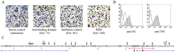
The cellular HERV-W envelope/syncytin-1 protein, encoded by the envelope gene of the ERVWE1 proviral locus is a fusogenic glycoprotein probably involved in the formation of the placental syncytiotrophoblast layer. Syncytin-1-induced in vitro cell-cell fusion is dependent on the interaction with hASCT2. As no receptor binding domain has been clearly defined in the SU of neither the HERV-W Env nor the retroviruses of the same interference group, we designed an in vitro binding assay to evaluate the interaction of the HERV-W envelope with the hASCT2 receptor. Using truncated HERV-W SU subunits, a region consisting of the N-terminal 124 amino acids of the mature SU glycoprotein was determined as the minimal receptor-binding domain. This domain contains several sub-domains which are poorly conserved among retroviruses of this interference group but a region of 18 residus containing the SDGGGX2DX2R conserved motif was proved to be essential for syncytin-1-hASCT2 interaction.


Figures

Figure 1
Figure 1
Cell surface binding assays of soluble SU. A) Schematic representation of HERV-W envelope protein (Env-W) and SU protein (EnvSU). Surface (SU, 1–313) and transmembrane (TM, 318–538) domains and the consensus furin cleavage site (RNKR, 314–317) are indicated. (|) N-glycosylation sites) [5]. Gray boxes indicate the signal peptide (SP) and a 15-amino-acid AAARVHRGS-H6 sequence (Tag). Residues are numbered starting from the initiation methionine. The ISKP sequence corresponds to the carboxy-terminal amino acid residues of the native SU included within the construct, and the P underlined amino acid corresponds to numbered residue located just upstream from the tag. B) Interaction of the soluble SU with the type D mammalian receptor. Soluble SU protein was secreted from HEK293T cells transfected with the SU domain expression vector and cultured for 24 h in medium without serum. Parental TE671, TE671 RD (hASCT2-blocked cells) and TE671 GALV (PiT1-blocked cells) cells were incubated at 37°C for 1 h in supernatants with (shaded) or without (white) soluble SU protein, collected from transfected or native HEK293T cells, respectively. Binding of the tagged soluble SU onto the cells was detected by incubating 1 h at 4°C the cell-protein mixture with an anti-histidine antibody (anti-RGS(H4)-Mab; Qiagen) in PBA (PBS with 2% fetal calf serum and 0.1% sodium azide). Cells were washed once and incubated with fluorescein isothiocyanate-conjugated antibody (DAKO) for 1 h at 4°C in PBA. The viable cells were analyzed by flow cytometry. C) Binding assay of SU soluble protein with target cells expressing various levels of hASCT1 and hASCT2 receptors. TE671, HeLa, HEK293T, XC-hASCT2 and XC-hASCT1 cells were incubated at 37°C for 1 h in HEK293T cell culture supernatants with (shaded) or without (white) soluble SU protein as indicated above. Binding assays were performed as described in 1B. D) Binding of SU soluble protein is restricted to human ASCT receptors. Soluble SU protein were incubated at 37°C for 1 h with parental XC rat cells (white) and XC-hASCT2 or XC-hASCT1 (shaded) stable cells Binding assays were performed as described in 1B.
Figure 2
Figure 2
Cell surface binding assays of SU and truncated proteins. A) Schematic representation of SU and truncated proteins. Gray boxes indicate signal peptide (SP) and 15-amino-acid AAARVHRGS-H6 sequence (Tag). Residues are numbered starting from the initiation methionine. ISKP, THTS, NFRP and VSLF sequences correspond to the carboxy-terminal amino acid residues of the native SU included within each construct, SU, Env233, Env197 and Env168, respectively. The underlined amino acids correspond to the numbered residue located just upstream from the tag. B) Detection of SU and truncated proteins in culture medium. Culture supernatants were collected from HEK293T cells transfected with either the SU domain or truncated envelope expression vectors as described in fig 1B. 20 μl of supernatant was denatured (0.5% sodium dodecyl sulfate [SDS], 1% β-mercaptoethanol) at 100°C for 10 min and analyzed by SDS-10% polyacrylamide gel electrophoresis. Blots were probed with with an anti-histidine antibody (anti-RGS(H4)-Mab; Qiagen). Blots were developed using horseradish peroxidase-conjugated antibodies (Jackson) together with an enhanced chemiluminescence kit (Amersham Pharmacia). * indicates the position of Env168 SU protein. C) Identification of the receptor binding domain. SU and truncated soluble proteins were incubated at 37°C for 1 h with XC-hASCT2 cells (shaded) or with parental XC cells (white). Binding assays were performed as described above. The binding capacity of each recombinant protein onto hASCT2 receptor is depicted with a green (efficient) or red (inefficient) highlighted name.
Figure 3
Figure 3
Validation of RBD functionality. A) Cell-cell fusion inhibition by the RBD. The wild-type HERV-W plasmid was transfected in TELCeB6 wild-type envelope producer cells phenotypically characterized by a nucleus expressing β-galactosidase. The HEK293T indicator target cells were transfected with antisense env gene (fusion control), Env71 (non-binding domain), Env144 (RBD) and EnvSU (inhibition control) plasmids. Producer cells were overlaid with HEK293T indicator cells transiently transfected with truncation mutants. Coculture were stained with X-Gal substrate then with May-Grünwald and Giemsa solutions. The determination of the fusion activity of the transfected envelope was performed after 20 h of coculture. The total number of syncytia in one well and the number of nuclei in each syncytia were determined. B) Binding inhibition by an anti-RBD antibody. The Env 144 protein was pre-incubated at 37°C for 1 h with the anti-SU and anti-TM antibodies (dotted line) [5]. Then, XC-hASCT2 cells were incubated at 37°C for 1 h with the protein-antibody complex. The shaded histogram (Env144 incubated with XC-hASCT2) and white histogram (Env144 incubated with parental XC cells) were indicated as controls. The binding was analyzed by flow cytometry with an anti-histidine antibody (anti-RGS(H4)-Mab; Qiagen). C) Receptor binding domain. Variable amino acids (aa) (green dot) among hominoids (upper-case), human (lower case) and neutral insertions preserving the envelope functions (white dots) are indicated above the sequence [23]. Strictly conserved aa (grey boxes) and similar aa (straight line) within the D interference group are underlined. Mutations altering spleen necrosis virus infectivity are indicated as black dots [24]. Blue arrows covering aa 21–69 and 117–144 indicate deletions detrimental to hASCT2 binding. The red arrow corresponds to the SU-EnvW peptide used for rabbit immunization and affinity purification [5].

Similar articles

Cited by

References

    1. Blond JL, Lavillette D, Cheynet V, Bouton O, Oriol G, Chapel-Fernandes S, Mandrand B, Mallet F, Cosset FL. An envelope glycoprotein of the human endogenous retrovirus HERV-W is expressed in the human placenta and fuses cells expressing the type D mammalian retrovirus receptor. J Virol. 2000;74:3321–3329. doi: 10.1128/JVI.74.7.3321-3329.2000. - DOI - PMC - PubMed
    1. Mi S, Lee X, Li X, Veldman GM, Finnerty H, Racie L, LaVallie E, Tang XY, Edouard P, Howes S, Keith JC, Jr, McCoy JM. Syncytin is a captive retroviral envelope protein involved in human placental morphogenesis. Nature. 2000;403:785–789. doi: 10.1038/35001608. - DOI - PubMed
    1. Frendo JL, Olivier D, Cheynet V, Blond JL, Bouton O, Vidaud M, Rabreau M, Evain-Brion D, Mallet F. Direct involvement of HERV-W Env glycoprotein in human trophoblast cell fusion and differentiation. Mol Cell Biol. 2003;23:3566–3574. doi: 10.1128/MCB.23.10.3566-3574.2003. - DOI - PMC - PubMed
    1. Mallet F, Bouton O, Prudhomme S, Cheynet V, Oriol G, Bonnaud B, Lucotte G, Duret L, Mandrand B. The endogenous retroviral locus ERVWE1 is a bona fide gene involved in hominoid placental physiology. Proc Natl Acad Sci USA. 2004;101:1731–1736. doi: 10.1073/pnas.0305763101. - DOI - PMC - PubMed
    1. Cheynet V, Ruggieri A, Oriol G, Blond JL, Boson B, Vachot L, Verrier B, Cosset FL, Mallet F. Synthesis, assembly, and processing of the Env ERVWE1/syncytin human endogenous retroviral envelope. J Virol. 2005;79:5585–5593. doi: 10.1128/JVI.79.9.5585-5593.2005. - DOI - PMC - PubMed

MeSH terms