

Phosphorylated epidermal growth factor receptor on tumor-associated endothelial cells in human renal cell carcinoma is a primary target for therapy by tyrosine kinase inhibitors
- PMID: 16820093
- PMCID: PMC1601465
- DOI: 10.1593/neo.06172
Phosphorylated epidermal growth factor receptor on tumor-associated endothelial cells in human renal cell carcinoma is a primary target for therapy by tyrosine kinase inhibitors
Abstract
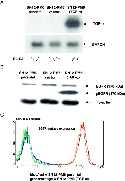
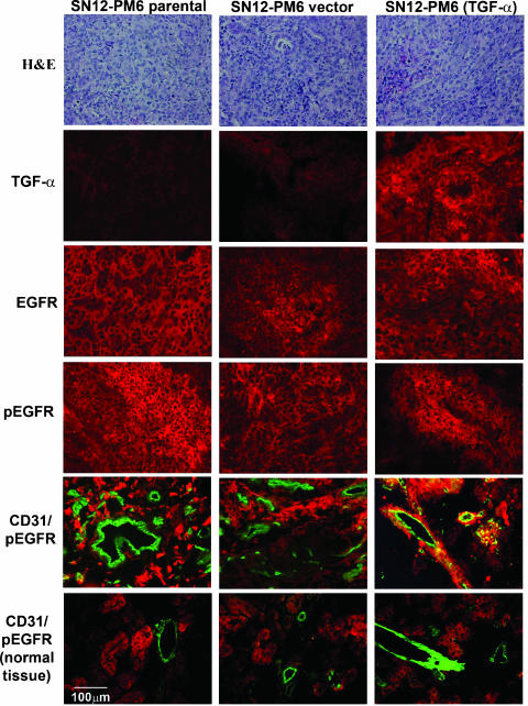
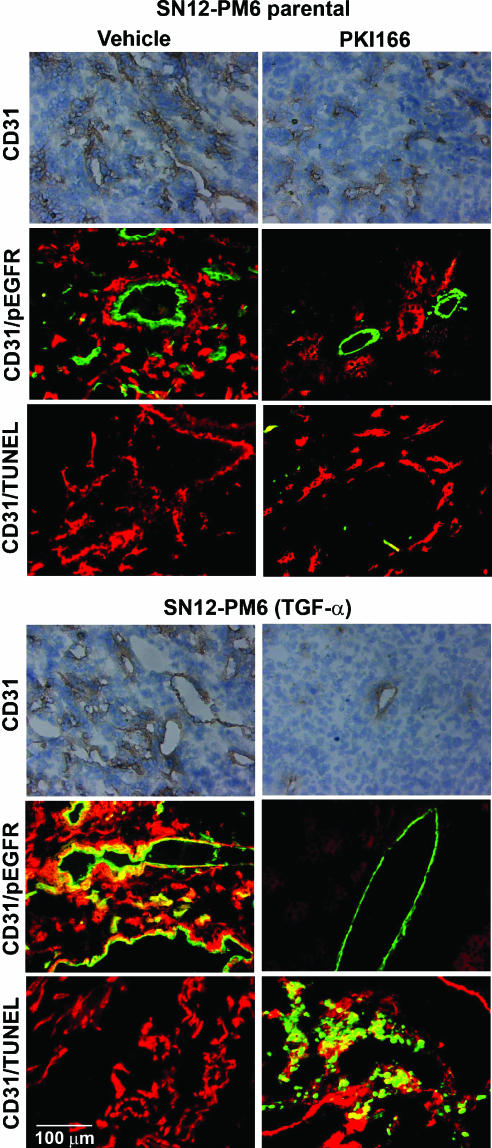
We determined whether therapy for human renal cell carcinoma (HRCC) that grows in the kidney of nude mice by the specific epidermal growth factor receptor (EGFR) tyrosine kinase inhibitor, PKI166, is directed against phosphorylated EGFR on tumor cells or on tumor-associated endothelial cells. EGFR+/transforming growth factor alpha (TGF-alpha)- SN12-PM6 HRCC cells were transfected with full-length sense TGF-alpha cDNA or vector control. SN12-PM6 cells expressing low or high levels of TGF-alpha were implanted into the kidney of nude mice. Only tumors produced by TGF-alpha+ HRCC cells contained tumor-associated endothelial cells expressing activated EGFR. Oral administration of PKI166 produced significant therapy only in TGF-alpha+ tumors, which correlated with apoptosis of tumor-associated endothelial cells. These data suggest that the production of TGF-alpha by HRCC cells leads to the activation of EGFR on tumor-associated endothelial cells that serve as an essential target for therapy with tyrosine kinase inhibitors.
Figures
Similar articles
-
Blockade of the epidermal growth factor receptor signaling inhibits angiogenesis leading to regression of human renal cell carcinoma growing orthotopically in nude mice.Clin Cancer Res. 2002 Nov;8(11):3592-600. Clin Cancer Res. 2002. PMID: 12429651
-
TGF-alpha-driven tumor growth is inhibited by an EGF receptor tyrosine kinase inhibitor.Biochem Biophys Res Commun. 2002 Jan 11;290(1):349-58. doi: 10.1006/bbrc.2001.6210. Biochem Biophys Res Commun. 2002. PMID: 11779176
-
Phosphorylated epidermal growth factor receptor on tumor-associated endothelial cells is a primary target for therapy with tyrosine kinase inhibitors.Neoplasia. 2008 May;10(5):489-500. doi: 10.1593/neo.08200. Neoplasia. 2008. PMID: 18472966 Free PMC article.
-
Critical update and emerging trends in epidermal growth factor receptor targeting in cancer.J Clin Oncol. 2005 Apr 10;23(11):2445-59. doi: 10.1200/JCO.2005.11.890. Epub 2005 Mar 7. J Clin Oncol. 2005. PMID: 15753456 Review.
-
[Recent advances in development of antitumor tyrosine kinase inhibitors].Gan To Kagaku Ryoho. 1997 Sep;24(11):1536-40. Gan To Kagaku Ryoho. 1997. PMID: 9309152 Review. Japanese.
Cited by
-
Neoplasia: the second decade.Neoplasia. 2008 Dec;10(12):1314-24. doi: 10.1593/neo.81372. Neoplasia. 2008. PMID: 19048110 Free PMC article.
-
Higher circulating expression levels of miR-221 associated with poor overall survival in renal cell carcinoma patients.Tumour Biol. 2014 May;35(5):4057-66. doi: 10.1007/s13277-013-1531-3. Epub 2013 Dec 31. Tumour Biol. 2014. PMID: 24379138
-
Signaling of ERBB receptor tyrosine kinases promotes neuroblastoma growth in vitro and in vivo.Cancer. 2010 Jul 1;116(13):3233-43. doi: 10.1002/cncr.25073. Cancer. 2010. PMID: 20564646 Free PMC article.
-
Inhibition of epidermal growth factor receptor and vascular endothelial growth factor receptor phosphorylation on tumor-associated endothelial cells leads to treatment of orthotopic human colon cancer in nude mice.Neoplasia. 2007 Dec;9(12):1066-77. doi: 10.1593/neo.07667. Neoplasia. 2007. PMID: 18084614 Free PMC article.
-
A Novel HER2 Protein Identification Methodology in Breast Cancer Cells Using Raman Spectroscopy and Raman Imaging: An Analytical Validation Study.J Med Chem. 2024 Oct 10;67(19):17629-17639. doi: 10.1021/acs.jmedchem.4c01591. Epub 2024 Sep 21. J Med Chem. 2024. PMID: 39305290 Free PMC article.
References
-
- Russo P. Renal cell carcinoma: presentation, staging, and surgical treatment. Semin Oncol. 2000;27:160–176. - PubMed
-
- Hartmann JT, Bokemeyer C. Chemotherapy for renal cell carcinoma. Anticancer Res. 1999;19:1541–1544. - PubMed
-
- Weber KL, Doucet M, Price JE, Baker CH, Kim SJ, Fidler IJ. Blockade of the epidermal growth factor receptor signaling leads to inhibition of renal cell carcinoma growth in the bone of nude mice. Cancer Res. 2003;63:2940–2947. - PubMed
-
- Kedar D, Baker CH, Killion JJ, Dinney CPN, Fidler IJ. Blockade of the epidermal growth factor receptor signaling inhibits angiogenesis leading to the regression of human renal cell carcinoma growing orthotopically in nude mice. Clin Cancer Res. 2002;8:3592–3600. - PubMed
-
- Traxler P, Buchdunger E, Furet P, Gschwind H-P, Ho P, Mett H. Preclinical profile of PKI166—a novel and potent EGFR tyrosine kinase inhibitor for clinical development. Clin Cancer Res. 1999;5:3750.
Publication types
MeSH terms
Substances
Grants and funding
LinkOut - more resources
Full Text Sources
Other Literature Sources
Research Materials
Miscellaneous