Review
. 2006 Nov;98(5):901-26.
doi: 10.1093/aob/mcl133. Epub 2006 Jul 4.

Structure-function relationships in highly modified shoots of cactaceae

Affiliations
Review

Structure-function relationships in highly modified shoots of cactaceae

James D Mauseth. Ann Bot. 2006 Nov.

Abstract

Background and aims: Cacti are extremely diverse structurally and ecologically, and so modified as to be intimidating to many biologists. Yet all have the same organization as most dicots, none differs fundamentally from Arabidopsis or other model plants. This review explains cactus shoot structure, discusses relationships between structure, ecology, development and evolution, and indicates areas where research on cacti is necessary to test general theories of morphogenesis.

Scope: Cactus leaves are diverse; all cacti have foliage leaves; many intermediate stages in evolutionary reduction of leaves are still present; floral shoots often have large, complex leaves whereas vegetative shoots have microscopic leaves. Spines are modified bud scales, some secrete sugar as extra-floral nectaries. Many cacti have juvenile/adult phases in which the flowering adult phase (a cephalium) differs greatly from the juvenile; in some, one side of a shoot becomes adult, all other sides continue to grow as the juvenile phase. Flowers are inverted: the exterior of a cactus 'flower' is a hollow vegetative shoot with internodes, nodes, leaves and spines, whereas floral organs occur inside, with petals physically above stamens. Many cacti have cortical bundles vascularizing the cortex, however broad it evolves to be, thus keeping surface tissues alive. Great width results in great weight of weak parenchymatous shoots, correlated with reduced branching. Reduced numbers of shoot apices is compensated by great increases in number of meristematic cells within individual SAMs. Ribs and tubercles allow shoots to swell without tearing during wet seasons. Shoot epidermis and cortex cells live and function for decades then convert to cork cambium. Many modifications permit water storage within cactus wood itself, adjacent to vessels.


Figures
F<sc>ig</sc>. 1
Fig. 1
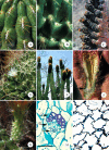
Cactus shoot structure. (A) Pereskia sacharosa with leaves. Stem about 6 mm across. (B) Maihuenia poeppigii, a geophyte; all green colour is due to small leaves. The yellow structure is a fruit. The entire plant is about 1 m across. (C) Growing cladode (long-shoot) of Opuntia violacea, with leaves still present (two indicated by arrows). Young cladode is emerging from an axillary bud of an older cladode. The spines of the axillary buds of the older cladode are bud scales. Slightly smaller than life size. (D). Shoot tip of Cereus forbesii. Three of four ribs are visible; spine clusters (axillary buds, areoles) are located along the rib apex. Spines are bud scales, dormant axillary bud shoot apical meristem is located just above each spine cluster, hidden by a mass of white trichomes. Spines are present even on very young axillary buds thus protecting the shoot apical meristem, which is not the highest point of the shoot (shoot apex is concave). Almost life size. (E) Old plant of Melocactus intortus with juvenile portion of shoot (green) produced during the first 10–15 years of the plant's life, and the adult portion (red, the cephalium), which is probably at least 10 years old. This is a single shoot (not a graft of two unrelated plants), produced by a single shoot apical meristem. No new chlorenchyma has been produced for years. (F). Shoots of Espostoa with lateral cephalia; flowers are produced only by axillary buds within the adult (cephalium) portion, not from the juvenile (green) portions. Note disrupted phyllotaxy. Shoots are about 5 cm across. (G) Areole (axillary bud) of Ferocactus. Spines are modified bud scales. The location of the bud apical meristem is indicated by an arrow, below the mass of trichomes. Trichomes (yellow) are abundant in the areole but absent from the rest of the shoot. Spines and trichomes emerge from a depression about 3 mm deep. About four times life size. (H) Leaves on floral bud of Browningia candelaris; the largest scale is about 1 cm across. Vegetative shoots of the same plant have only microscopic foliage leaves. (I) Longitudinal section of fully developed foliage leaf (L) of Oreocereus trollii; present are epidermis, stomata (not visible here), vascular tissue, chlorenchyma, dorsiventral asymmetry. Leaf is 450 μm tall. Axillary bud SAM (out of view on left) has produced leaf primordia, one of which is developing as a spine (S). Cells in spine base are meristematic, those in upper portion are elongating into fibres. Scale bar = 300 μm.
F<sc>ig</sc>. 2
Fig. 2
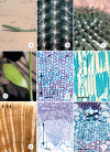
Specialized features of cacti. (A) Elongated axillary buds of Oroya peruviana, with spines in two rows, not in spiral phyllotaxy around the bud SAM, which is located at the top of each areole. Each areole is about 5 mm long. (B) Tubercles of Coryphantha clavata, each with an elongate areole containing one or two secretory spines (arrow). Note ordinary non-secretory spines at the tips of each tubercle (out of focus in foreground). Tubercles are about 12 mm long. (C) Elongated areoles of Neoraimondia roseiflora. When first formed, these resembled ordinary areoles as in Fig. 1D, but each has flowered numerous times over many years, growing longer each time. Each has bark, cortex, stele and pith. These are about 60 mm long. (D) Dimorphic areoles of Mammillaria camptotricha. Each areole SAM has divided into two, one being carried outward with the tubercle apex where it makes only spines, the other remaining at the tubercle base where it produces a flower or a vegetative shoot. The open flower is about 8 mm across. (E) Terminal cephalia of a single, branched plant of Backebergia militaris. Green portions are juvenile, cephalia are the adult body. Each shoot tip will be abscised about 4 cm below the cephalium, then one or two axillary buds will grow as new juvenile bodies for several years, then convert to making cephalia. The plant is about 6 m tall. (F) Section of a floral shoot of Neocardenasia. The outer portion is a long-shoot with leaves, nodes and internodes. True flower organs occur only along the inner surface (upper arrow indicates boundary between vegetative and floral organs); petals and stamens, although located physically above the ovary and style base, are morphologically lower (proximal). After fertilization, all tissues above the lower arrow will abscise, removing style, stamens, petals and much vegetative tissue. The region below the lower arrow will develop into a true fruit surrounded by a false fruit. (G) External structure of floral shoot of Echinocereus. Although referred to as a ‘flower,’ this is long-shoot tissue with tiny foliage leaves, axillary buds (bud scales are spines), nodes and internodes. True floral structures are present inside this shoot (some petal bases are visible at the top). Almost life size. (H) Cortical bundle in Lepismium, with xylem (x), phloem (ph) and a cap of phloem fibres (f). All conducting cells are extremely narrow. Scale bar = 100 μm. (I) Collapsible cortex in Haageocereus. Completely turgid palisade cortex cells are <500 μm away in the same region. Scale bar = 100 μm.
F<sc>ig</sc>. 3
Fig. 3
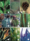
Cactus structures. (A) Prostrate shoot of Harrisia pomanensis. The shoot tip is elevated by reaction cortex near the phloem on the lower side of the shoot. The visible part of the shoot is about 40 cm long; the entire shoot is several metres long, branched and growing in various directions. The lower side has adventitious roots. (B) Three ribs of a columnar cactus, Coleocephalocereus. As the shoot loses water and volume, ribs become narrower but do not change surface area. Each spine cluster is an axillary bud; subtending foliage leaves (like those in Fig. 1I) are microscopic. Each areole could potentially produce a vegetative long-shoot (a branch); because this species has lateral cephalia, these are juvenile phase areoles and cannot bloom. Each rib base is about 10 mm across. (C) Tubercles of Mammillaria magnimamma, produced in obvious phyllotactic spirals. Tuberculate shoots shrink vertically as well as radially as water is lost. Being a Mammillaria, this has divided, dimorphic areoles: areole SAMs at tubercle tips produce only spines; areole SAMs at tubercle bases (hidden by white trichomes) produce floral shoots or vegetative shoots (none are present in the photograph). Each tubercle is about 10 mm long. (D) Dimorphic shoots of Epiphyllum caudatum; the branch on the left was initially terete but immediately switched to distichous phyllotaxy with just two tall, thin ribs. The ‘midvein’ is the central vascular cylinder, the ‘blade’ is the two ribs and notches along the rib crests are the axillary buds (spines are microscopic). The vertical shoot on the right is terete here, but its tip had also switched to growing as a two-ribbed leaf-like structure. The cladode is about 30 mm across. (E) Wide-band tracheid wood of Thelocactus bicolor in transverse section. The double-headed arrow in the lower portion indicates the region with many vessels (dark red, narrower walls), perhaps earlywood. The upper portion of the micrograph is mostly WBTs (perhaps latewood), with two vessels (arrows). In many WBTs, the band-like secondary wall almost occludes the lumen. Scale bar = 100 μm. (F) Tangential section of WBT wood in Thelocactus; WBTs are short and imperforate, and in this species the secondary wall occurs as one or two helices per cell. Blue is the flexible primary wall; despite the thick secondary wall, these cells shorten and lengthen as the water content changes. Cells near the left, lacking wide-bands, are ray parenchyma cells. Scale bar = 100 μm. (G) Transverse view (macroscopic) of Consolea wood. Rays (white, arrowed) are very wide (1 to 3 mm); vessels within the axial masses (tan) are close to water stored in the rays. These rays interconnect water stored in the pith and cortex. The image is about 30 mm across. (H) Dimorphic wood of Stenocereus. The double-headed arrow indicates primary xylem and first-formed secondary xylem, both lacking fibres and instead having WBTs, vessels and xylem parenchyma. After several months, the vascular cambium switched to making fibrous wood (above upper arrowhead), consisting of vessels, xylem parenchyma and xylary fibres but no WBTs. Scale bar = 100 μm. (I) Transverse section of secondary phloem of Corryocactus. The arrow indicates collapsed phloem, below which are abundant sieve tube members and companion cells. A phloem fibre cap is at the top of the image, secondary xylem at the bottom. Scale bar = 100 μm.

References

    1. Anderson EF. (2001) The cactus family(Timber Press, Portland, OR).
    1. Arias S, Terrazas T, Cameron K. (2003) Phylogenetic analysis of Pachycereus (Cactaceae, Pachycereeae) based on chloroplast and nuclear DNA sequences. Systematic Botany 28547–557.
    1. Arias Montes AS. (1996) Revisión taxonómica del género Pereskiopsis Britton et Rose (Cactaceae). (Universidad Nacional Autónoma de México, Mexico).
    1. Arnold DH and Mauseth JD. (1999) Effects of environmental factors on development of wood. American Journal of Botany 86367–371. - PubMed
    1. Backeberg C. (1958) Die Cactaceae. Handbuch der Kakteenkunde. I. Einleitung und Beschreibung der Peireskioideae und Opuntioideae(VEB Gustav Fisher Verlag, Jena).

Publication types