. 2006 Jul 4;34(11):3288-98.
doi: 10.1093/nar/gkl401. Print 2006.

SAP30L interacts with members of the Sin3A corepressor complex and targets Sin3A to the nucleolus

Affiliations

SAP30L interacts with members of the Sin3A corepressor complex and targets Sin3A to the nucleolus

K M Viiri et al. Nucleic Acids Res. .

Abstract

Histone acetylation plays a key role in the regulation of gene expression. The chromatin structure and accessibility of genes to transcription factors is regulated by enzymes that acetylate and deacetylate histones. The Sin3A corepressor complex recruits histone deacetylases and in many cases represses transcription. Here, we report that SAP30L, a close homolog of Sin3-associated protein 30 (SAP30), interacts with several components of the Sin3A corepressor complex. We show that it binds to the PAH3/HID (Paired Amphipathic Helix 3/Histone deacetylase Interacting Domain) region of mouse Sin3A with residues 120-140 in the C-terminal part of the protein. We provide evidence that SAP30L induces transcriptional repression, possibly via recruitment of Sin3A and histone deacetylases. Finally, we characterize a functional nucleolar localization signal in SAP30L and show that SAP30L and SAP30 are able to target Sin3A to the nucleolus.


Figures
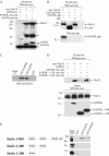
Figure 1
Figure 1
SAP30L interacts with Sin3A. (A) Sin3A co-immunoprecipitates with SAP30L. Lysates from the transfected HEK293T cells were immunoprecipitated with agarose conjugated anti-his antibody, and the immunocomplexes were analyzed by western blotting with anti-myc antibody. (B) SAP30L co-immunoprecipitates with the Sin3A protein. Transfected HEK293T cells were lysed and immunoprecipitated with the agarose conjugated anti-myc antibody, and the immunocomplexes were analyzed by western blotting with anti-myc and anti-gfp antibodies. (C) GST-SAP30L and GST-SAP30 pulls down endogenous Sin3A protein from nuclear lysate of HEK293T cells, whereas GST alone does not. Western blotting was performed with Sin3A antibody. (D) Residues between 120 and 140 in the SAP30L protein are critical for the association with the Sin3A. Immunocomplexes in the upper panel and inputs in the lower panel were analyzed by western blotting with anti-myc antibody. (E) SAP30L binds directly to PAH3/HID domain in Sin3A. Sin3A constructs used are illustrated on the left side of the experimental panel. The Sin3A proteins were produced by coupled in vitro transcription/translation system and labeled with 35S-Methionine before subjecting to pull-down experiments with GST-fusion proteins as indicated. SDS–PAGE was subjected to autoradiography. (Asterisk, IgG heavy and light chains; GST, glutathione-S-transferase; PAH, paired amhipathic helix; HID, histone deacetylase interacting domain.)
Figure 2
Figure 2
SAP30L is able to self-associate. (A) Myc-his-tagged SAP30L associates with gfp-tagged SAP30L in vivo. Lysates from the transfected HEK293T cells were immunoprecipitated with agarose conjugated anti-myc antibody and the immunocomplexes were analyzed with the antibodies indicated. (B) GST-SAP30L can directly interact with in vitro translated SAP30L. SAP30L proteins were produced by coupled in vitro transcription/translation and labeled with 35S-methionine and subjected to pull-down experiment with full-length GST-SAP30L as indicated. Pull-down complexes were analyzed by autoradiography. The amount of bound protein was quantified and normalized to the input. Data are illustrated in the histogram. The mutants of SAP30L are shown below.
Figure 3
Figure 3
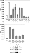
SAP30L associates with histone deacetylase activity. (A and B) GST-fusion pull-downs from the HEK293T nuclear extracts were performed and HDAC activities were measured with the Fluor de Lys kit. Schematic representation of SAP30L constructs used are shown in Figures 2b and 5. GST and GST-SAP30 were used as a negative and positive control, respectively. The basal level of fluorescence (blank) and sensitivity in the experiment were defined by measuring fluorescence from the assay buffer and from the 1 µM deacetylated standard respectively (white bars). NAD+ coenzyme and TSA were added in the cases indicated. Shown are the means of two experiments performed in duplicate and the error bars represent the range of measurements (AFU = arbitrary fluorescence unit). (C) GST-SAP30 and GST-SAP30L pulls down endogenous HDAC 1 and 2, and transfected myc-his tagged HDAC 3 from the HEK293T lysates, whereas GST alone does not (negative control). Pull-down complexes were analyzed by western blotting with the antibodies indicated.
Figure 4
Figure 4
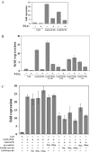
SAP30L is able to repress transcription. (AC) HEK293T cells were cotransfected with 5xGal4-14D luciferase reporter vector, Gal4DBD fusions and LacZ-vector as indicated. Twenty-four hour post-transfection cells were either treated with TSA or DMSO (vehicle) for another 24 h. Lysed cells were analyzed for luciferase and β-gal activity. The histogram illustrates the average fold-repressions of the Gal4DBD-fusions compared with Gal4 alone. The measurements were done in duplicate from two independent experiments and the error bars represent the range.
Figure 5
Figure 5
SAP30L localizes in the nucleolus. (A), SAP30L colocalizes with nucleophosmin (NPM) in the nucleolus. Ten hour treatment with proteasome inhibitor MG132 causes further accumulation of SAP30L to the nucleolus. (B) NoLS signal resides between residues 120–140 in SAP30L. Arrows indicate cytoplasmic accumulation of the SAP30L 1–120 and SAP30L-NLSmut mutants. Arrowhead indicates the nucleolus. (C) Residues 120–127 in the SAP30L protein are necessary for its nucleolar accumulation. In the above experiments, HEK293T cells were transfected with the indicated myc-his-tagged constructs, and double-stained with anti-myc (construct) and anti-nucleophosmin (nucleolar marker) antibodies. Cells were further stained with DAPI in order to visualize the nuclei. Cells in the right panel were treated for 10 h with 10 µM proteasome inhibitor MG132. All the pictures were taken by confocal fluorescence microscope. Line-diagrams illustrate the fluorescence intensity (green, SAP30L; red, NPM) along the white line shown in merged images. SAP30L-myc-his constructs used are illustrated on the left side of each panel. (D) The NoLS of SAP30L and SAP30 identified in this study was manually aligned with three other previously published nucleolus localizations sequences of the following human proteins: catalytic subunit of human telomerase [TERT (34)]; NOLP (35) and hLa (21).
Figure 6
Figure 6
SAP30 and SAP30L target Sin3A to the nucleolus. (A) Cotransfected SAP proteins target Sin3A to the nucleolus. Myc-Sin3A was transfected with either myc-his tagged SAP30L, SAP30L 1–120 or SAP30 and cells were treated for 10 h with MG132. Sin3A protein was visualized by the Sin3A antibody and SAP proteins with the His-tag antibody. Stained cells were analyzed with confocal microscopy. Arrows indicate the nucleoli. (B) Data in A (without MG132) were scored as percent of cells expressing Sin3A in the nucleoli (total 100 cells counted for each experiment) and illustrated as the histogram.
Figure 7
Figure 7
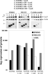
Fast turnover of the SAP30L protein correlates with its nucleolar localization. HEK293T cells were cotransfected with the indicated, myc-his-tagged constructs and a control vector (LacZ-myc-his). Cells were treated 10 h with either DMSO (vehicle), MG132 (proteasome inhibitor) or cyclohexamide (translation inhibitor). The lysed cells were subjected to SDS–PAGE and analyzed by Western blotting with anti-myc and anti-actin (loading control) antibodies. Band intensities were quantified and the amount of the protein in the sample was calculated after normalization to LacZ expression. Representative results from three independent experiments are shown.

Similar articles

Cited by

References

    1. Ayer D.E. Histone deacetylases: transcriptional repression with SINers and NuRDs. Trends. Cell Biol. 1999;9:193–198. - PubMed
    1. Narlikar G.J., Fan H.Y., Kingston R.E. Cooperation between complexes that regulate chromatin structure and transcription. Cell. 2002;108:475–487. - PubMed
    1. Santoro R., Grummt I. Epigenetic mechanism of rRNA gene silencing: temporal order of NoRC-mediated histone modification, chromatin remodeling, and DNA methylation. Mol. Cell Biol. 2005;25:2539–2546. - PMC - PubMed
    1. Silverstein R.A., Ekwall K. Sin3: a flexible regulator of global gene expression and genome stability. Curr. Genet. 2005;47:1–17. - PubMed
    1. Zhang Y., Iratni R., Erdjument-Bromage H., Tempst P., Reinberg D. Histone deacetylases and SAP18, a novel polypeptide, are components of a human Sin3 complex. Cell. 1997;89:357–364. - PubMed

Publication types