

Mutagenic nucleotide incorporation and hindered translocation by a food carcinogen C8-dG adduct in Sulfolobus solfataricus P2 DNA polymerase IV (Dpo4): modeling and dynamics studies
- PMID: 16820532
- PMCID: PMC1500869
- DOI: 10.1093/nar/gkl425
Mutagenic nucleotide incorporation and hindered translocation by a food carcinogen C8-dG adduct in Sulfolobus solfataricus P2 DNA polymerase IV (Dpo4): modeling and dynamics studies
Abstract
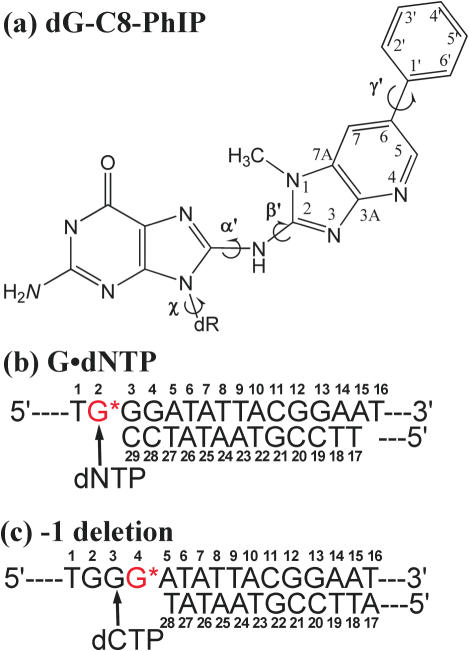
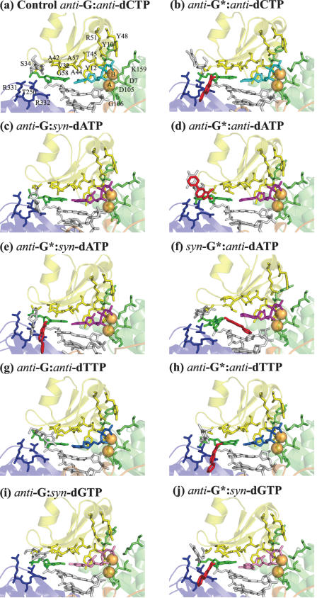
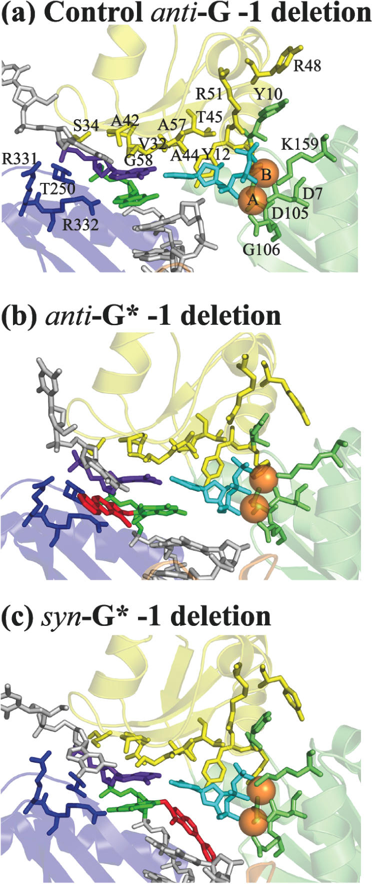
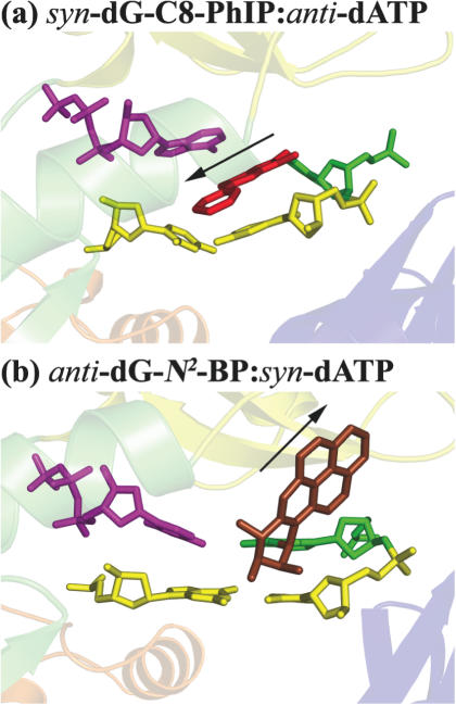
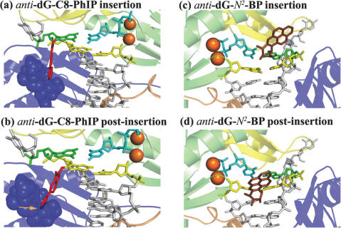
Bulky carcinogen-DNA adducts commonly cause replicative polymerases to stall, leading to a switch to bypass polymerases. We have investigated nucleotide incorporation opposite the major adduct of 2-amino-1-methyl-6-phenylimidazo[4,5-b]pyridine (PhIP) in the DinB family polymerase, Dpo4, using molecular modeling and molecular dynamics (MD) simulations. PhIP, the most prevalent heterocyclic aromatic amine formed by cooking of proteinaceous food, is mutagenic in mammalian cells and is implicated in mammary and colon tumors. Our results show that the dG-C8-PhIP adduct can be accommodated in the spacious major groove Dpo4 open pocket, with Dpo4 capable of incorporating dCTP, dTTP or dATP opposite the adduct reasonably well. However, the PhIP ring system on the minor groove side would seriously disturb the active site, regardless of the presence and identity of dNTP. Furthermore, the simulations indicate that dATP and dTTP are better incorporated in the damaged system than in their respective mismatched but unmodified controls, suggesting that the PhIP adduct enhances incorporation of these mismatches. Finally, bulky C8-dG adducts, situated in the major groove, are likely to impede translocation in this polymerase (Rechkoblit et al. (2006), PLoS Biol., 4, e11). However, N2-dG adducts, which can reside on the minor groove side, appear to cause less hindrance when in this position.
Figures
Similar articles
-
How Y-Family DNA polymerase IV is more accurate than Dpo4 at dCTP insertion opposite an N2-dG adduct of benzo[a]pyrene.DNA Repair (Amst). 2015 Nov;35:144-53. doi: 10.1016/j.dnarep.2015.09.020. Epub 2015 Sep 26. DNA Repair (Amst). 2015. PMID: 26523515
-
A new anti conformation for N-(deoxyguanosin-8-yl)-2-acetylaminofluorene (AAF-dG) allows Watson-Crick pairing in the Sulfolobus solfataricus P2 DNA polymerase IV (Dpo4).Nucleic Acids Res. 2006 Feb 1;34(3):785-95. doi: 10.1093/nar/gkj479. Print 2006. Nucleic Acids Res. 2006. PMID: 16452300 Free PMC article.
-
Molecular Insights into the Translesion Synthesis of Benzyl-Guanine from Molecular Dynamics Simulations: Structural Evidence of Mutagenic and Nonmutagenic Replication.Biochemistry. 2017 Apr 4;56(13):1841-1853. doi: 10.1021/acs.biochem.6b01247. Epub 2017 Mar 24. Biochemistry. 2017. PMID: 28290677
-
Toward an understanding of the role of DNA adduct conformation in defining mutagenic mechanism based on studies of the major adduct (formed at N(2)-dG) of the potent environmental carcinogen, benzo[a]pyrene.Mutat Res. 2000 May 30;450(1-2):41-59. doi: 10.1016/s0027-5107(00)00015-4. Mutat Res. 2000. PMID: 10838133 Review.
-
Mutagenicity of Ochratoxin A: Role for a Carbon-Linked C8-Deoxyguanosine Adduct?J Agric Food Chem. 2017 Aug 23;65(33):7097-7105. doi: 10.1021/acs.jafc.6b03897. Epub 2016 Nov 15. J Agric Food Chem. 2017. PMID: 28830149 Review.
Cited by
-
DNA adduct structure-function relationships: comparing solution with polymerase structures.Chem Res Toxicol. 2008 Jan;21(1):45-52. doi: 10.1021/tx700193x. Epub 2007 Dec 4. Chem Res Toxicol. 2008. PMID: 18052109 Free PMC article. Review.
-
Effect of oxidatively damaged DNA on the active site preorganization during nucleotide incorporation in a high fidelity polymerase from Bacillus stearothermophilus.Proteins. 2008 May 15;71(3):1360-72. doi: 10.1002/prot.21824. Proteins. 2008. PMID: 18058909 Free PMC article.
-
p53 triggers mitochondrial apoptosis following DNA damage-dependent replication stress by the hepatotoxin methyleugenol.Cell Death Dis. 2022 Nov 29;13(11):1009. doi: 10.1038/s41419-022-05446-9. Cell Death Dis. 2022. PMID: 36446765 Free PMC article.
-
A Unique B-Family DNA Polymerase Facilitating Error-Prone DNA Damage Tolerance in Crenarchaeota.Front Microbiol. 2020 Jul 23;11:1585. doi: 10.3389/fmicb.2020.01585. eCollection 2020. Front Microbiol. 2020. PMID: 32793138 Free PMC article.
-
Preferred WMSA catalytic mechanism of the nucleotidyl transfer reaction in human DNA polymerase κ elucidates error-free bypass of a bulky DNA lesion.Nucleic Acids Res. 2012 Oct;40(18):9193-205. doi: 10.1093/nar/gks653. Epub 2012 Jul 5. Nucleic Acids Res. 2012. PMID: 22772988 Free PMC article.
References
-
- Lehmann A.R. Replication of damaged DNA by translesion synthesis in human cells. FEBS Lett. 2005;579:873–876. - PubMed
-
- Prakash S., Johnson R.E., Prakash L. Eukaryotic translesion synthesis DNA polymerases: specificity of structure and function. Annu. Rev. Biochem. 2005;74:317–353. - PubMed
-
- Wagner J., Gruz P., Kim S.R., Yamada M., Matsui K., Fuchs R.P., Nohmi T. The dinB gene encodes a novel E.coli DNA polymerase, DNA pol IV, involved in mutagenesis. Mol. Cell. 1999;4:281–286. - PubMed
Publication types
MeSH terms
Substances
Grants and funding
LinkOut - more resources
Full Text Sources
Other Literature Sources
Miscellaneous
