

Cloning, overexpression, purification and preliminary crystallographic studies of a mitochondrial type II peroxiredoxin from Pisum sativum
- PMID: 16820697
- PMCID: PMC2242941
- DOI: 10.1107/S1744309106023451
Cloning, overexpression, purification and preliminary crystallographic studies of a mitochondrial type II peroxiredoxin from Pisum sativum
Abstract
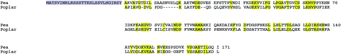
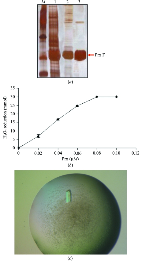
A cDNA encoding an open reading frame of 199 amino acids corresponding to a type II peroxiredoxin from Pisum sativum with its transit peptide was isolated by RT-PCR. The 171-amino-acid mature protein (estimated molecular weight 18.6 kDa) was cloned into the pET3d vector and overexpressed in Escherichia coli. The recombinant protein was purified and crystallized by the hanging-drop vapour-diffusion technique. A full data set (98.2% completeness) was collected using a rotating-anode generator to a resolution of 2.8 angstroms from a single crystal flash-cooled at 100 K. X-ray data revealed that the protein crystallizes in space group P1, with unit-cell parameters a = 61.88, b = 66.40, c = 77.23 angstroms, alpha = 102.90, beta = 104.40, gamma = 99.07 degrees, and molecular replacement using a theoretical model predicted from the primary structure as a search model confirmed the presence of six molecules in the unit cell as expected from the Matthews coefficient. Refinement of the structure is in progress.
Figures
Similar articles
-
Crystallization and preliminary X-ray data of a bifunctional peroxiredoxin from poplar.Acta Crystallogr D Biol Crystallogr. 2002 Sep;58(Pt 9):1501-3. doi: 10.1107/S0907444902011782. Epub 2002 Aug 23. Acta Crystallogr D Biol Crystallogr. 2002. PMID: 12198315
-
Crystallization and preliminary X-ray diffraction analysis of thioredoxin peroxidase from the aerobic hyperthermophilic archaeon Aeropyrum pernix K1.Acta Crystallogr Sect F Struct Biol Cryst Commun. 2005 Mar 1;61(Pt 3):323-5. doi: 10.1107/S1744309105005294. Epub 2005 Feb 24. Acta Crystallogr Sect F Struct Biol Cryst Commun. 2005. PMID: 16511031 Free PMC article.
-
Purification, crystallization and preliminary crystallographic study of a recombinant plant aminoaldehyde dehydrogenase from Pisum sativum.Acta Crystallogr Sect F Struct Biol Cryst Commun. 2008 Feb 1;64(Pt 2):88-90. doi: 10.1107/S1744309107068522. Epub 2008 Jan 18. Acta Crystallogr Sect F Struct Biol Cryst Commun. 2008. PMID: 18259056 Free PMC article.
-
Crystallization and preliminary X-ray analysis of Escherichia coli p20, a novel thiol peroxidase.Acta Crystallogr D Biol Crystallogr. 2003 Jun;59(Pt 6):1064-6. doi: 10.1107/s0907444903006930. Epub 2003 May 23. Acta Crystallogr D Biol Crystallogr. 2003. PMID: 12777775
-
Cloning and characterization of a 2-Cys peroxiredoxin from Pisum sativum.J Exp Bot. 2004 Oct;55(406):2191-9. doi: 10.1093/jxb/erh238. Epub 2004 Aug 27. J Exp Bot. 2004. PMID: 15333640
Cited by
-
X-ray crystallographic and high-speed AFM studies of peroxiredoxin 1 from Chlamydomonas reinhardtii.Acta Crystallogr F Struct Biol Commun. 2018 Feb 1;74(Pt 2):86-91. doi: 10.1107/S2053230X17018507. Epub 2018 Jan 26. Acta Crystallogr F Struct Biol Commun. 2018. PMID: 29400317 Free PMC article.
-
Dissecting the integrative antioxidant and redox systems in plant mitochondria. Effect of stress and S-nitrosylation.Front Plant Sci. 2013 Nov 28;4:460. doi: 10.3389/fpls.2013.00460. eCollection 2013. Front Plant Sci. 2013. PMID: 24348485 Free PMC article.
-
The dual-targeted plant sulfiredoxin retroreduces the sulfinic form of atypical mitochondrial peroxiredoxin.Plant Physiol. 2011 Feb;155(2):944-55. doi: 10.1104/pp.110.166504. Epub 2010 Dec 7. Plant Physiol. 2011. PMID: 21139087 Free PMC article.
-
Calcium sensing via EF-hand 4 enables thioredoxin activity in the sensor-responder protein calredoxin in the green alga Chlamydomonas reinhardtii.J Biol Chem. 2020 Jan 3;295(1):170-180. doi: 10.1074/jbc.RA119.008735. Epub 2019 Nov 27. J Biol Chem. 2020. PMID: 31776187 Free PMC article.
-
Peroxiredoxins: a less studied component of hydrogen peroxide detoxification in photosynthetic organisms.Protoplasma. 2009 Mar;235(1-4):3-15. doi: 10.1007/s00709-009-0032-0. Epub 2009 Feb 15. Protoplasma. 2009. PMID: 19219525 Review.
References
-
- Alphey, M. S., Bond, C. S., Tetaud, E., Fairlamb, A. H. & Hunter, W. N. (2000). J. Mol. Biol.300, 903–916. - PubMed
-
- Bernier-Villamor, L., Navarro, E., Sevilla, F. & Lázaro, J. J. (2004). J. Exp. Bot.55, 2191–2199. - PubMed
-
- Dietz, K. J. (2003). Annu. Rev. Plant Physiol. Plant Mol. Biol.54, 93–107. - PubMed
-
- Echalier, A., Trivelli, X., Corbier, C., Rouhier, N., Walker, O., Tsan, P., Jacquot, J.-P., Aubry, A., Krimm, I. & Lancelin, J.-M. (2005). Biochemistry, 44, 1755–1767. - PubMed
-
- Finkemeier, I., Goodman, M., Lankemeyer, P., Kandlbinder, A., Sweetlove, L. J. & Dietz, K. J. (2005). J. Biol. Chem.280, 12168–12180. - PubMed
Publication types
MeSH terms
Substances
LinkOut - more resources
Full Text Sources
