

Contrast enhancement in dense breast images to aid clustered microcalcifications detection
- PMID: 16820957
- PMCID: PMC3043882
- DOI: 10.1007/s10278-005-6976-5
Contrast enhancement in dense breast images to aid clustered microcalcifications detection
Abstract
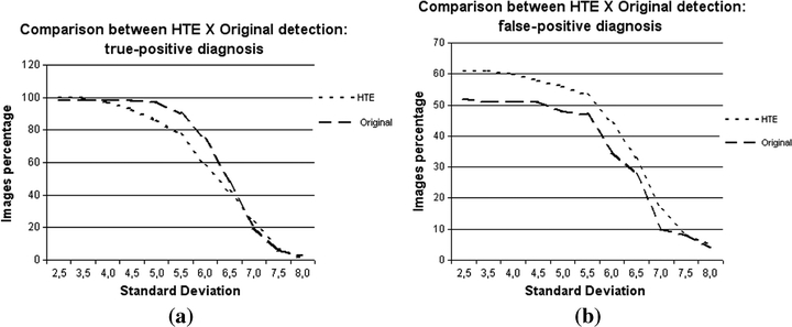
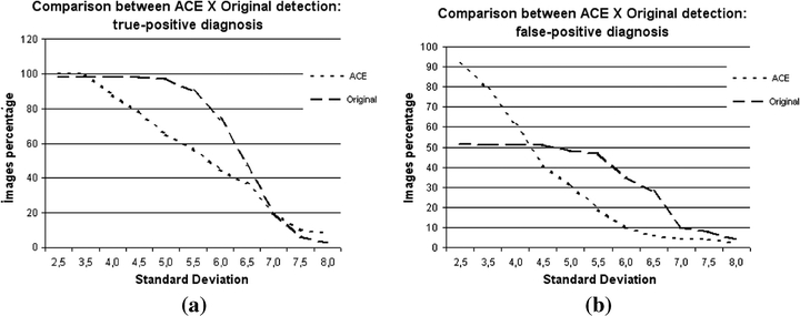
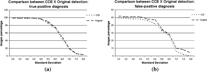
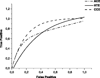
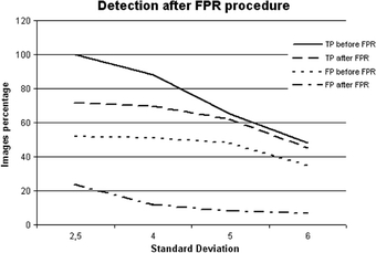
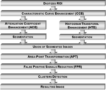
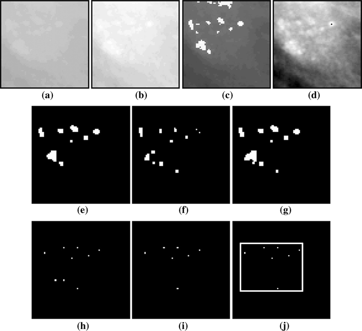
This paper presents a method to provide contrast enhancement in dense breast digitized images, which are difficult cases in testing of computer-aided diagnosis (CAD) schemes. Three techniques were developed, and data from each method were combined to provide a better result in relation to detection of clustered microcalcifications. Results obtained during the tests indicated that, by combining all the developed techniques, it is possible to improve the performance of a processing scheme designed to detect microcalcification clusters. It also allows operators to distinguish some of these structures in low-contrast images, which were not detected via conventional processing before the contrast enhancement. This investigation shows the possibility of improving CAD schemes for better detection of microcalcifications in dense breast images.
Figures

Similar articles
-
Segmentation of suspicious clustered microcalcifications in mammograms.Med Phys. 2000 Jan;27(1):13-22. doi: 10.1118/1.598852. Med Phys. 2000. PMID: 10659733
-
Computer-aided detection of clustered microcalcifications in digitized mammograms.Acad Radiol. 1995 Aug;2(8):655-62. doi: 10.1016/s1076-6332(05)80431-3. Acad Radiol. 1995. PMID: 9419620
-
Computerized detection of clustered microcalcifications in digital mammograms using a shift-invariant artificial neural network.Med Phys. 1994 Apr;21(4):517-24. doi: 10.1118/1.597177. Med Phys. 1994. PMID: 8058017
-
Detection of potential microcalcification clusters using multivendor for-presentation digital mammograms for short-term breast cancer risk estimation.Med Phys. 2019 Apr;46(4):1938-1946. doi: 10.1002/mp.13450. Epub 2019 Mar 7. Med Phys. 2019. PMID: 30801718 Free PMC article. Review.
-
Computer-aided detection and diagnosis of breast cancer with mammography: recent advances.IEEE Trans Inf Technol Biomed. 2009 Mar;13(2):236-51. doi: 10.1109/TITB.2008.2009441. Epub 2009 Jan 20. IEEE Trans Inf Technol Biomed. 2009. PMID: 19171527 Review.
Cited by
-
Foundation and methodologies in computer-aided diagnosis systems for breast cancer detection.EXCLI J. 2017 Feb 20;16:113-137. doi: 10.17179/excli2016-701. eCollection 2017. EXCLI J. 2017. PMID: 28435432 Free PMC article. Review.
-
A new fast fractal modeling approach for the detection of microcalcifications in mammograms.J Digit Imaging. 2010 Oct;23(5):538-46. doi: 10.1007/s10278-009-9224-6. Epub 2009 Jul 18. J Digit Imaging. 2010. PMID: 19618243 Free PMC article.
-
Methods Used in Computer-Aided Diagnosis for Breast Cancer Detection Using Mammograms: A Review.J Healthc Eng. 2020 Mar 12;2020:9162464. doi: 10.1155/2020/9162464. eCollection 2020. J Healthc Eng. 2020. PMID: 32300474 Free PMC article. Review.
-
Mammographic image denoising and enhancement using the Anscombe transformation, adaptive wiener filtering, and the modulation transfer function.J Digit Imaging. 2013 Apr;26(2):183-97. doi: 10.1007/s10278-012-9507-1. J Digit Imaging. 2013. PMID: 22806627 Free PMC article.
References
-
- Giger ML. Computer-aided diagnosis of breast lesions in medical images. Comput Sci Eng. 2000;2(5):39–45. doi: 10.1109/5992.877391. - DOI
-
- Giger ML, MacMahon H. Image processing and computer-aided diagnosis. Radiol Clin North Am. 1996;34(3):565–595. - PubMed
-
- LeGal M, Chavanne G, Pellier D. Valeur diagnostique des microcalcifications groupées découvertes par mammographies. Bull Cancer. 1984;71:57–64. - PubMed
-
- Nishikawa RM, Jiang Y, Giger ML, Schimdt RA, Vyborny CJ, Zhang W, Papaionnou J, Bick U, Nagel R, Doi K, et al. Performance of automated CAD schemes for the detection and classification of clustered microcalcifications. In: Gale AG, et al., editors. Digital Mammography. Amsterdam: Elsevier; 1994. pp. 13–20.
-
- Roerih J, et al.: Clinical results with R2 imagechecker system. In: Karssemeijer N, et al. (Eds). Digital Mammography. Kluwer Academic Publishing, Dordrecht, The Netherlands, 1998, pp 395–400
Publication types
MeSH terms
Substances
LinkOut - more resources
Full Text Sources
Medical
Miscellaneous
