Post-embryonic remodelling of neurocranial elements: a comparative study of normal versus abnormal eye migration in a flatfish, the Atlantic halibut
- PMID: 16822267
- PMCID: PMC2100306
- DOI: 10.1111/j.1469-7580.2006.00577.x
Post-embryonic remodelling of neurocranial elements: a comparative study of normal versus abnormal eye migration in a flatfish, the Atlantic halibut
Abstract
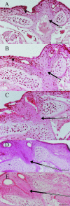
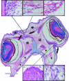
The process of eye migration in bilaterally symmetrical flatfish larvae starts with asymmetrical growth of the dorsomedial parts of the ethmoid plate together with the frontal bones, structures initially found in a symmetrical position between the eyes. The movement of these structures in the future ocular direction exerts a stretch on the fibroblasts in the connective tissue found between the moving structures and the eye that is to migrate. Secondarily, a dense cell population of fibroblasts ventral to the eye starts to proliferate, possibly cued by the pulling forces exerted by the eye. The increased growth ventral to the eye pushes the eye dorsally. Osteoblasts are deposited in the dense cell layer, forming the dermal part of the lateral ethmoid, and at full eye migration this will cover the area vacated by the migrated eye. When the migrating eye catches up with the previous migrated dermal bones, the frontals, these bones will be remodelled to accommodate the eye. Our findings suggest that a combination of extremely localized signals and more distant factors may impinge upon the outcome of the tissue remodelling. Early normal asymmetry of signalling factors may cascade on a series of events.
Figures


Similar articles
-
Twisted story of eye migration in flatfish.J Morphol. 2006 Jun;267(6):730-8. doi: 10.1002/jmor.10437. J Morphol. 2006. PMID: 16526052
-
Distortion of frontal bones results from cell apoptosis by the mechanical force from the up-migrating eye during metamorphosis in Paralichthys olivaceus.Mech Dev. 2015 May;136:87-98. doi: 10.1016/j.mod.2015.01.001. Epub 2015 Jan 23. Mech Dev. 2015. PMID: 25622577
-
Proliferating cells in suborbital tissue drive eye migration in flatfish.Dev Biol. 2011 Mar 1;351(1):200-7. doi: 10.1016/j.ydbio.2010.12.032. Epub 2010 Dec 31. Dev Biol. 2011. PMID: 21195706
-
Flatfish: an asymmetric perspective on metamorphosis.Curr Top Dev Biol. 2013;103:167-94. doi: 10.1016/B978-0-12-385979-2.00006-X. Curr Top Dev Biol. 2013. PMID: 23347519 Review.
-
Orchestrating change: The thyroid hormones and GI-tract development in flatfish metamorphosis.Gen Comp Endocrinol. 2015 Sep 1;220:2-12. doi: 10.1016/j.ygcen.2014.06.012. Epub 2014 Jun 26. Gen Comp Endocrinol. 2015. PMID: 24975541 Review.
Cited by
-
A thyroid hormone regulated asymmetric responsive centre is correlated with eye migration during flatfish metamorphosis.Sci Rep. 2018 Aug 16;8(1):12267. doi: 10.1038/s41598-018-29957-8. Sci Rep. 2018. PMID: 30115956 Free PMC article.
-
Teleost Metamorphosis: The Role of Thyroid Hormone.Front Endocrinol (Lausanne). 2019 Jun 14;10:383. doi: 10.3389/fendo.2019.00383. eCollection 2019. Front Endocrinol (Lausanne). 2019. PMID: 31258515 Free PMC article. Review.
-
Iodine and selenium supplementation increased survival and changed thyroid hormone status in Senegalese sole (Solea senegalensis) larvae reared in a recirculation system.Fish Physiol Biochem. 2012 Jun;38(3):725-34. doi: 10.1007/s10695-011-9554-4. Epub 2011 Sep 20. Fish Physiol Biochem. 2012. PMID: 21932022
-
Unraveling the transcriptomic landscape of eye migration and visual adaptations during flatfish metamorphosis.Commun Biol. 2024 Mar 1;7(1):253. doi: 10.1038/s42003-024-05951-x. Commun Biol. 2024. PMID: 38429383 Free PMC article.
References
-
- Benjamin M. Mucochondroid (mucous connective) tissues in the heads of teleosts. Anat Embryol. 1988;178:461–474. - PubMed
-
- Bjornsson BT, Halldorsson O, Haux C, Norberg B, Brown CL. Photoperiod control of sexual maturation of the Atlantic halibut (Hippoglossus hippoglossus): plasma thyroid hormone calcium levels. Aquaculture. 1998;166:117–140.
-
- Boyle WJ, Simonet WS, Lacey DL. Osteoclast differentiation and activation. Nature. 2003;423:337–342. - PubMed
-
- Brewster B. Eye migration and cranial development during flatfish metamorphosis: a reappraisal (teleostei: Pleuronectiformes) J Fish Biol. 1987;31:805–833.
-
- Hagiwara H, Inoue A, Nakajo S, et al. Inhibition of proliferation of chondrocytes by specific receptors in response to retinoids. Biochem Biophys Res Commun. 1996;222:220–224. - PubMed
Publication types
MeSH terms
Substances
LinkOut - more resources
Full Text Sources
Medical

