

Expression of pituitary adenylate cyclase-activating polypeptide in the primary lymphoid organs of the duck Anas platyrhynchos
- PMID: 16822269
- PMCID: PMC2100303
- DOI: 10.1111/j.1469-7580.2006.00592.x
Expression of pituitary adenylate cyclase-activating polypeptide in the primary lymphoid organs of the duck Anas platyrhynchos
Abstract
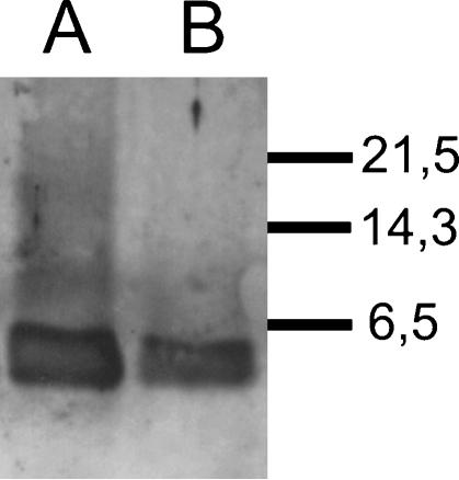
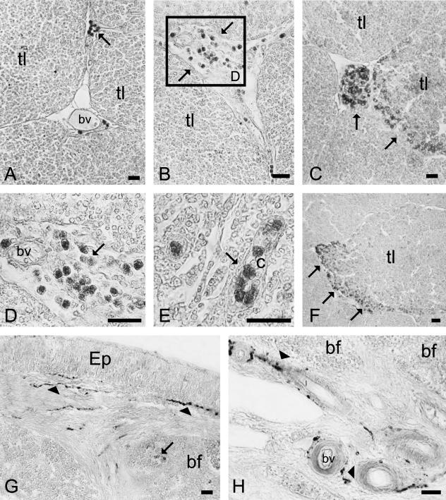
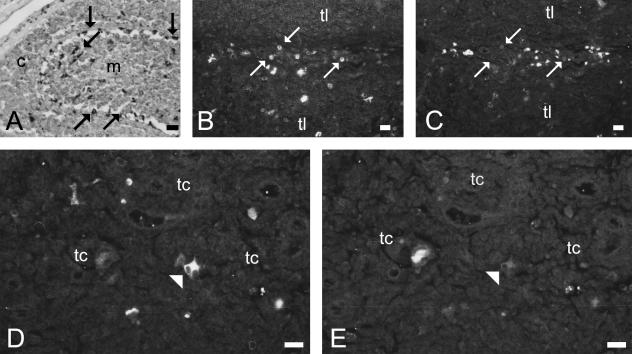
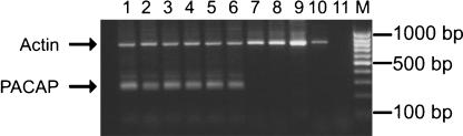
The expression of pituitary adenylate cyclase-activating polypeptide (PACAP) was studied in the thymus and bursa of Fabricius of the duck Anas platyrhynchos, at different ages, using immunohistochemistry, Western blotting, RT-PCR and sequencing. In the thymus, PACAP immunoreactivity (-ir) was found in lymphoid cells. CD68/ and PGP 9.5/PACAP38 double labelling showed that PACAP was not expressed either in macrophages or in epithelial cells, suggesting that the PACAP-positive cells observed were lymphoid cells. Immunoreactive lymphocytes were observed in the interlobular septa. They increased in number with ageing. In the bursa, PACAP-ir was found in nerve fibres and in a few lymphoid cells. RT-PCR revealed PACAP mRNA expression in the thymus but not in the bursa. These results suggest that PACAP plays a role in the functions of the immune system in birds.
Figures
Similar articles
-
Pituitary adenylyl cyclase-activating polypeptide (PACAP) and its receptor (PAC1-R) in the cochlea: evidence for specific transcript expression of PAC1-R splice variants in rat microdissected cochlear subfractions.Neuroscience. 2006 Jun 19;140(1):147-61. doi: 10.1016/j.neuroscience.2006.01.019. Epub 2006 Apr 19. Neuroscience. 2006. PMID: 16626868
-
Pituitary adenylate cyclase activating peptide (PACAP) immunoreactivity and mRNA expression in the duck gastrointestinal tract.Cell Tissue Res. 2002 Jun;308(3):347-59. doi: 10.1007/s00441-002-0555-6. Epub 2002 Apr 26. Cell Tissue Res. 2002. PMID: 12107428
-
Expression localisation and functional activity of pituitary adenylate cyclase-activating polypeptide, vasoactive intestinal polypeptide and their receptors in mouse ovary.Reproduction. 2007 Aug;134(2):281-92. doi: 10.1530/REP-07-0051. Reproduction. 2007. PMID: 17660238
-
The presence and distribution of pituitary adenylate cyclase activating polypeptide and its receptor in the snail Helix pomatia.Neuroscience. 2008 Aug 13;155(2):387-402. doi: 10.1016/j.neuroscience.2008.05.003. Epub 2008 May 8. Neuroscience. 2008. PMID: 18590802
-
Pituitary adenylate cyclase-activating polypeptide and PACAP receptor expression and function in the rat adrenal gland.Int J Mol Med. 2002 Mar;9(3):233-43. Int J Mol Med. 2002. PMID: 11836629
Cited by
-
Relationship between Plasma Pituitary Adenylate Cyclase-Activating Polypeptide (PACAP) Level and Proteome Profile of Cows.Animals (Basel). 2022 Jun 16;12(12):1559. doi: 10.3390/ani12121559. Animals (Basel). 2022. PMID: 35739894 Free PMC article.
-
Distribution and molecular evolution of the neuropeptide pituitary adenylate cyclase-activating polypeptide (PACAP) and its receptors in the lizard Podarcis sicula (Squamata, Lacertidae).J Mol Neurosci. 2009 Sep;39(1-2):144-56. doi: 10.1007/s12031-009-9178-7. Epub 2009 Jan 28. J Mol Neurosci. 2009. PMID: 19184550
-
Immunomodulatory Role of Neuropeptides in the Cornea.Biomedicines. 2022 Aug 16;10(8):1985. doi: 10.3390/biomedicines10081985. Biomedicines. 2022. PMID: 36009532 Free PMC article. Review.
References
-
- Abad C, Martinez C, Leceta J, Juarranz MG, Delgado M, Gomariz RP. Pituitary adenylate-cyclase-activating polypeptide expression in the immune system. Neuroimmunomodulation. 2002;10:177–186. - PubMed
-
- Ali SM, Chan AS, Leong SK. Histochemical and immunohistochemical localization of nitrergic neuronal and non-neuronal cells in the bursa of Fabricius of the chicken. Cell Tissue Res. 1996;285:273–279. - PubMed
-
- Batanero E, De Leeuw FE, Hansen GH, Van Wichen DF, Huber J, Schuurman HJ. The neural and neuro-endocrine component of the human thymus. II. Hormone immunoreactivity. Brain Behav Immun. 1992;6:249–264. - PubMed
-
- Bishop CM, McCabe CJ, Gittoes NJL, Butler PJ, Franklyn JA. Tissue-specific regulation of thyroid hormone receptor mRNA isoforms and target gene proteins in domestic ducks. J Endocrinol. 2000;165:507–615. - PubMed
MeSH terms
Substances
LinkOut - more resources
Full Text Sources
Medical
Miscellaneous
