Four-bar linkage modelling in teleost pharyngeal jaws: computer simulations of bite kinetics
- PMID: 16822272
- PMCID: PMC2100308
- DOI: 10.1111/j.1469-7580.2006.00551.x
Four-bar linkage modelling in teleost pharyngeal jaws: computer simulations of bite kinetics
Abstract
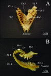
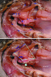
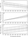
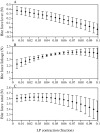
The pharyngeal arches of the red drum (Sciaenops ocellatus) possess large toothplates and a complex musculoskeletal design for biting and crushing hard prey. The morphology of the pharyngeal apparatus is described from dissections of six specimens, with a focus on the geometric conformation of contractile and rotational elements. Four major muscles operate the rotational 4th epibranchial (EB4) and 3rd pharyngobranchial (PB3) elements to create pharyngeal bite force, including the levator posterior (LP), levator externus 3/4 (LE), obliquus posterior (OP) and 3rd obliquus dorsalis (OD). A biomechanical model of upper pharyngeal jaw biting is developed using lever mechanics and four-bar linkage theory from mechanical engineering. A pharyngeal four-bar linkage is proposed that involves the posterior skull as the fixed link, the LP muscle as input link, the epibranchial bone as coupler link and the toothed pharyngobranchial as output link. We used a computer model to simulate contraction of the four major muscles, with the LP as the dominant muscle, the length of which determined the position of the linkage. When modelling lever mechanics, we found that the effective mechanical advantages of the pharyngeal elements were low, resulting in little resultant bite force. By contrast, the force advantage of the four-bar linkage was relatively high, transmitting approximately 50% of the total muscle force to the bite between the toothplates. Pharyngeal linkage modelling enables quantitative functional morphometry of a key component of the fish feeding system, and the model is now available for ontogenetic and comparative analyses of fishes with pharyngeal linkage mechanisms.
Figures




Similar articles
-
Functional morphology of the pharyngeal jaw apparatus in perciform fishes: An experimental analysis of the haemulidae.J Morphol. 1989 Jun;200(3):231-245. doi: 10.1002/jmor.1052000302. J Morphol. 1989. PMID: 29865647
-
A biomechanical model for analysis of muscle force, power output and lower jaw motion in fishes.J Theor Biol. 2003 Aug 7;223(3):269-81. doi: 10.1016/s0022-5193(03)00058-4. J Theor Biol. 2003. PMID: 12850448
-
Functional morphology of the pharyngeal jaw apparatus in moray eels.J Morphol. 2008 May;269(5):604-19. doi: 10.1002/jmor.10612. J Morphol. 2008. PMID: 18196573
-
Masticatory muscles and the skull: a comparative perspective.Arch Oral Biol. 2007 Apr;52(4):296-9. doi: 10.1016/j.archoralbio.2006.09.010. Epub 2006 Nov 7. Arch Oral Biol. 2007. PMID: 17084804 Free PMC article. Review.
-
Biomechanics of occlusion--implications for oral rehabilitation.J Oral Rehabil. 2016 Mar;43(3):205-14. doi: 10.1111/joor.12345. Epub 2015 Sep 15. J Oral Rehabil. 2016. PMID: 26371622 Review.
Cited by
-
The morphology of the mouse masticatory musculature.J Anat. 2013 Jul;223(1):46-60. doi: 10.1111/joa.12059. Epub 2013 May 20. J Anat. 2013. PMID: 23692055 Free PMC article.
-
High-performance suction feeding in an early elasmobranch.Sci Adv. 2019 Sep 11;5(9):eaax2742. doi: 10.1126/sciadv.aax2742. eCollection 2019 Sep. Sci Adv. 2019. PMID: 31535026 Free PMC article.
-
Phenotypic covariation predicts diversification in an adaptive radiation of pupfishes.Ecol Evol. 2024 Aug 7;14(8):e11642. doi: 10.1002/ece3.11642. eCollection 2024 Aug. Ecol Evol. 2024. PMID: 39114171 Free PMC article.
-
Form and function of damselfish skulls: rapid and repeated evolution into a limited number of trophic niches.BMC Evol Biol. 2009 Jan 30;9:24. doi: 10.1186/1471-2148-9-24. BMC Evol Biol. 2009. PMID: 19183467 Free PMC article.
References
-
- Aerts P, Verraes W. Theoretical analysis of a planar four-bar linkage in the teleostean skull. The use of mathematics in biomechanics. Ann Soc Roy Zool Belg. 1984;114:273–290.
-
- Alfaro ME, Bolnick DI, Wainwright PC. Evolutionary dynamics of complex biomechanical systems: an example using the four-bar mechanism. Evolution. 2004;58:495–503. - PubMed
-
- Anker G. Morphology and kinetics of the stickleback, Gasterosteus aculeatus. Trans Zool Soc Lond. 1974;32:311–416.
-
- Barel CDN, van der Meulen JW, Berkhoudt H. Kinematischer transmissionskoeffizient und vierstangensystem als funktionsparameter und formmodell für mandibular depressionsapparate bei Teleostiern. Anat Anz. 1977;142:21–31. - PubMed
-
- Boothby RN, Avault JW. Food habits, length–weight relationship, and condition factor of the red drum (Sciaenops ocellata) in Southeastern Louisiana. Trans Amer Fish Soc. 1971;2:1–18.
Publication types
MeSH terms
LinkOut - more resources
Full Text Sources
Miscellaneous

