

Microcracks in compact bone: a three-dimensional view
- PMID: 16822275
- PMCID: PMC2100300
- DOI: 10.1111/j.1469-7580.2006.00554.x
Microcracks in compact bone: a three-dimensional view
Abstract
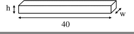
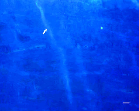
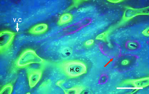
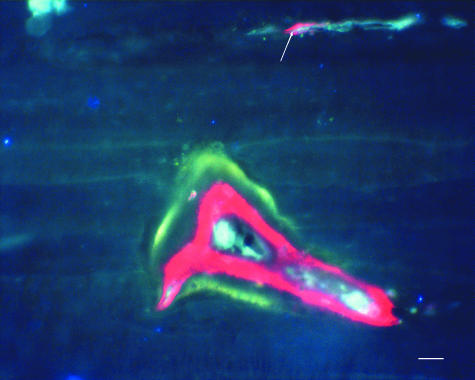
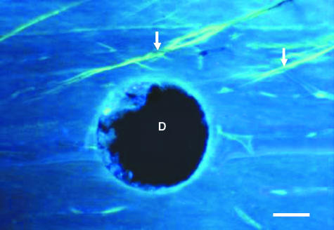
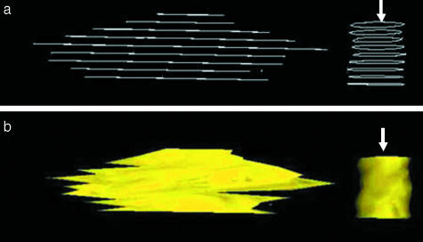
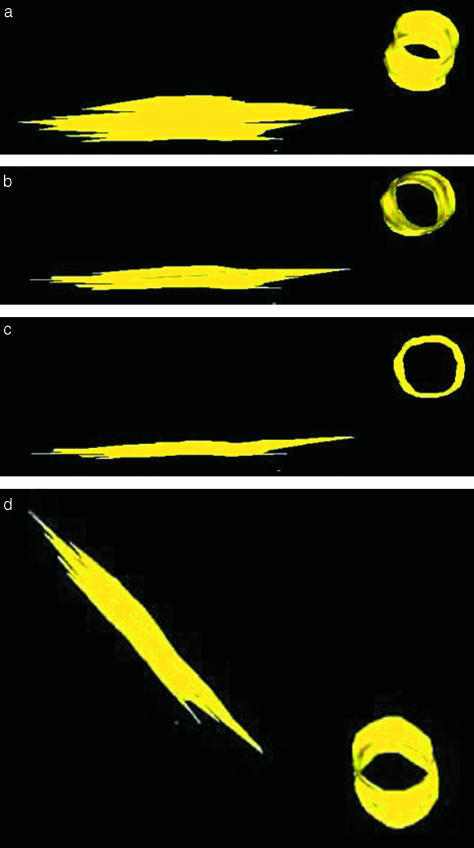
Microcracks have been implicated in the loss of bone quality for osteoporosis. In order to detect and monitor their growth, and to use these data to predict fractures, it is essential to obtain quantitative data regarding their shape in three dimensions. Beam-shaped bone samples from sheep radii were prepared and stained with fluorochrome dyes and tested in cyclical fatigue under four-point bending in a servo-hydraulic fatigue-testing machine. Samples were tested at a frequency of 30 Hz under load control at a stress range of 100 MPa. Holes were drilled into bone samples and used as reference points for reconstructions. A series of thin longitudinal sections were cut using a sledge macrotome. A two-dimensional image of each section was examined using an epifluorescence microscope and images transferred to a PC via a CCD low-light colour video camera. A three-dimensional image of each microcrack was reconstructed using computer software, and its dimensions measured. Cracks were elliptical in shape, longer in the longitudinal direction and with a mean aspect ratio of 5.5 +/- 1.05. The mean (+/- SD) length and width of labelled microcracks were 488 +/- 151 and 88 +/- 21 microm, respectively.
Figures
Similar articles
-
Three-dimensional reconstruction of Haversian systems in ovine compact bone.Eur J Morphol. 2002 Dec;40(5):309-15. doi: 10.1076/ejom.40.5.309.28901. Eur J Morphol. 2002. PMID: 15101447
-
Visualisation of three-dimensional microcracks in compact bone.J Anat. 2000 Oct;197 Pt 3(Pt 3):413-20. doi: 10.1046/j.1469-7580.2000.19730413.x. J Anat. 2000. PMID: 11117627 Free PMC article.
-
Osteonal crack barriers in ovine compact bone.J Anat. 2006 Jan;208(1):81-9. doi: 10.1111/j.1469-7580.2006.00509.x. J Anat. 2006. PMID: 16420381 Free PMC article.
-
Micro-tomographic imaging for the nondestructive evaluation of trabecular bone architecture.Stud Health Technol Inform. 1997;40:61-79. Stud Health Technol Inform. 1997. PMID: 10168883 Review.
-
The cellular transducer in bone: What is it?Technol Health Care. 2006;14(4-5):367-77. Technol Health Care. 2006. PMID: 17065758 Review.
Cited by
-
Measurement of the mechanical properties of bone: a recent history.Clin Orthop Relat Res. 2009 Aug;467(8):1948-54. doi: 10.1007/s11999-009-0784-z. Epub 2009 Mar 14. Clin Orthop Relat Res. 2009. PMID: 19288162 Free PMC article.
-
Long-term effects of bisphosphonate therapy: perforations, microcracks and mechanical properties.Sci Rep. 2017 Mar 6;7:43399. doi: 10.1038/srep43399. Sci Rep. 2017. PMID: 28262693 Free PMC article.
-
Detecting stress injury (fatigue fracture) in fibular cortical bone using quantitative ultrashort echo time-magnetization transfer (UTE-MT): An ex vivo study.NMR Biomed. 2018 Nov;31(11):e3994. doi: 10.1002/nbm.3994. Epub 2018 Jul 30. NMR Biomed. 2018. PMID: 30059184 Free PMC article.
-
Synchrotron radiation micro-CT at the micrometer scale for the analysis of the three-dimensional morphology of microcracks in human trabecular bone.PLoS One. 2011;6(7):e21297. doi: 10.1371/journal.pone.0021297. Epub 2011 Jul 7. PLoS One. 2011. PMID: 21750707 Free PMC article.
-
Aging and bone.J Dent Res. 2010 Dec;89(12):1333-48. doi: 10.1177/0022034510377791. Epub 2010 Oct 5. J Dent Res. 2010. PMID: 20924069 Free PMC article. Review.
References
-
- Bentolila V, Boyce TM, Fyhrie DP, et al. Intracortical remodeling in adult rat long bones after fatigue loading. Bone. 1998;23:275–281. - PubMed
-
- Bouxsein ML. Bone quality: where do we go from here? Osteoporos Int. 2003;14(Suppl. 5):118–127. - PubMed
-
- Burr DB, Martin RB, Schaffler MB, Radin EL. Bone remodeling in response to in vivo fatigue microdamage. J Biomech. 1985;18:189–200. - PubMed
-
- Burr DB, Milgrom C, Boyd RD, Higgins WL, Robin G, Radin EL. Experimental stress fractures of the tibia. Biological and mechanical aetiology in rabbits. J Bone Joint Surg. 1990;72B:370–375. - PubMed
Publication types
MeSH terms
LinkOut - more resources
Full Text Sources
Medical
