Comparative Study
. 2006 Aug 30;1107(1):70-81.
doi: 10.1016/j.brainres.2006.05.102. Epub 2006 Jul 5.

Spatiotemporal evolution of apoptotic neurodegeneration following traumatic injury to the developing rat brain

Affiliations
Comparative Study

Spatiotemporal evolution of apoptotic neurodegeneration following traumatic injury to the developing rat brain

Philip V Bayly et al. Brain Res. .

Abstract

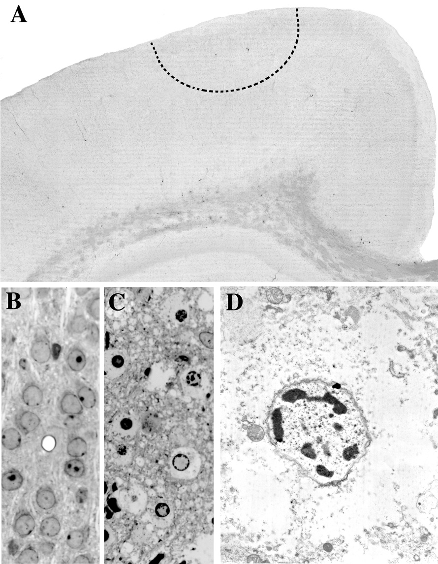
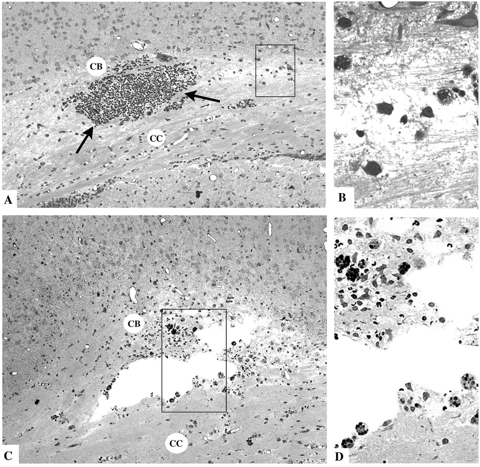
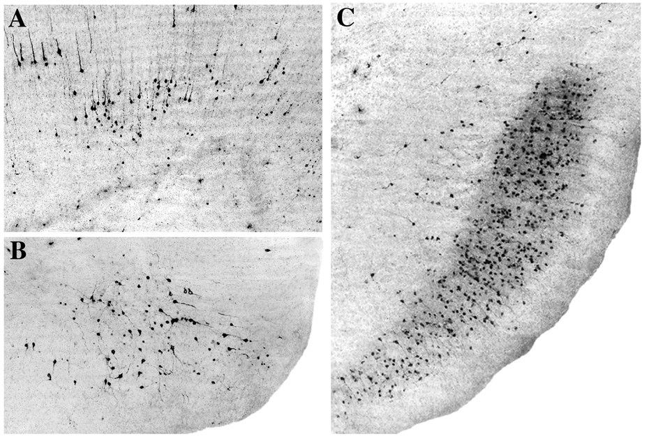
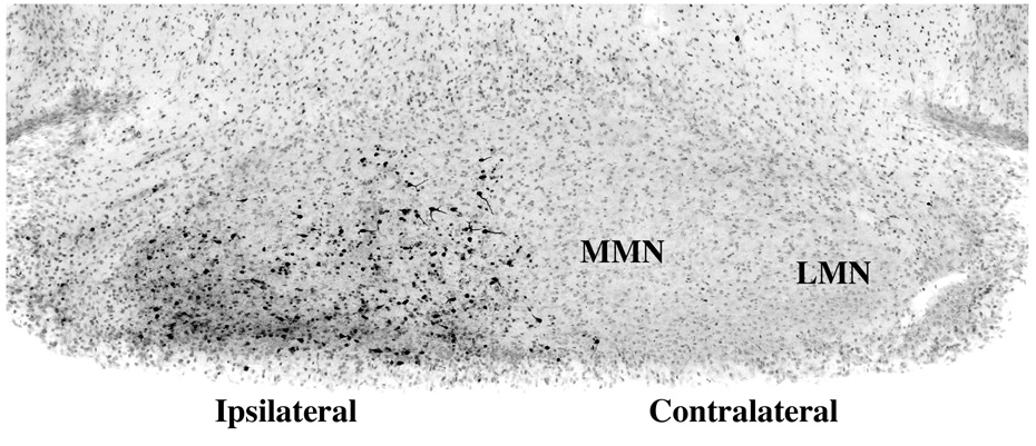
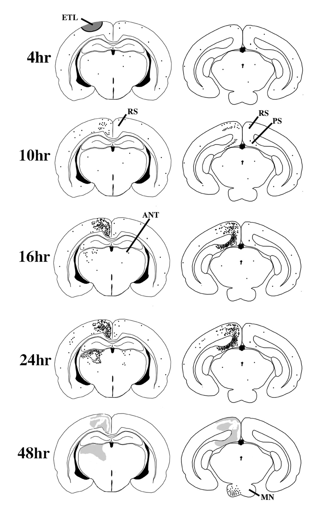
Closed head injury to the developing rat brain causes an acute excitotoxic lesion and axonal disruption at the impact site followed by a delayed pattern of apoptotic damage at various distant sites. Using an electromagnetic impact device to deliver a precisely controlled degree of mechanical deformation to the P7 infant rat skull, we studied the distribution of distant apoptotic lesions and the sequence and time course with which these lesions evolve following relatively mild closed head injury. The first major wave of apoptotic neurodegeneration occurred at 8 h postimpact in the retrosplenial cortex and pre- and parasubiculum. The next major wave occurred in the 16- to 24-h interval and was localized to the anterior thalamic nuclei. A third wave was detected at 36 to 48 h in the mammillary nuclei. We propose that the first and second waves were triggered by injury to a specific fiber tract, the corpus callosum/cingulum bundle that conveys reciprocal connections between the anterior thalamic nuclei and retrosplenial/pre- and parasubicular neurons. This fiber tract passes through a zone of maximum mechanical strain, as measured by tagged MRI. The third wave affecting mammillary neurons occurred because the principal synaptic targets of these neurons are the anterior thalamic neurons that were destroyed in the second wave of degeneration. Prevention of these apoptotic waves of brain damage is a realistic goal in view of the long delay between the impact event and onset of apoptotic degeneration.


Figures

Fig. 1
Fig. 1
Panel A is a 50 µm thick AC-3 stained vibratome section from a P7 rat brain 4 hrs post-impact. An acute excitotoxic neurodegenerative reaction is occurring at the site of impact within the region demarcated by the dashed semi-circular line, but this is not detected by the AC-3 stain because the excitotoxic lesion induced by head trauma does not entail caspase-3 activation. Panels B and C are 1 µm thin plastic sections from the same brain region demarcated by the dashed line in A depicting the appearance of normal neurons in a control brain (B) and abnormal appearance of neurons undergoing acute excitotoxic degeneration in an experimental brain 4 hrs post-impact (C). Panel D is an electron micrograph showing the ultrastructural appearance of a neuron (from the scene in C) undergoing excitotoxic cell death. These cells display all the morphological changes characteristic of an excitotoxic process. In the initial stages, dendrites and cell bodies undergo extreme edematous swelling which is accompanied by dilatation of mitochondria and endoplasmic reticulum, disaggregation of polyribosomes and dissolution of the formed structural elements in the cytoplasm. Nuclear changes occur slightly later and begin by the formation of small aggregates of clumped chromatin which give the nucleus a floccular appearance. These small clumps migrate to the perimeter of the nucleus in a clockface pattern, then coalesce into progressively larger clumps that consolidate into a large irregular dense mass at the center of the nucleus (nuclear pyknosis). The nuclear membrane remains intact but the plasma membrane becomes ruptured and loses its integrity early in the degenerative process. Compare this description with that given for neuroapoptosis in the legend of Fig. 7.
Fig. 2
Fig. 2
Thin plastic sections from the corpus callosum (CC) and cingulum bundle (CB) region of the infant rat brain 4 hours (A and B) or 16 hours (C and D) after TBI. The typical findings at 4 hours are a mild degree of interstitial edema giving portions of the CCCB track a rarefied appearance, and focal accumulations of red blood cells (arrows) due apparently to rupture of small blood vessels. The boxed region in A, shown at higher magnification in B, exhibits axonal fibers in disarray, surrounded by edema fluid and accompanied by phagocytic cells that are just beginning to respond to the pathological reaction. At 16 hrs, cystic spaces have formed at the site of tissue injury. The boxed regions in C, shown at higher magnification in D, reveals many phagocytic cells lining the cystic spaces and occupying the surrounding tissue area where they are ingesting red blood cells (dark round masses) and other less dense debris.
Fig. 3
Fig. 3
All panels are AC-3 stained sections from the P7 rat brain showing the first wave of caspase-3 activation that occurs at distant sites on a delayed basis following brain impact. The earliest AC-3 response occurs at 8–10 hrs and selectively affects the retrosplenial cortex (A) and parasubiculum (B). Initially, a relatively small number of neurons are involved, but in the ensuing 6 hours many more neurons in these same locations become involved. For example, panel C shows the extensive involvement of many neurons in the pre and parasubicular region 16 hrs following impact, compared to the relatively small patch of neurons involved at 8–10 hrs (panel B).
Fig. 4
Fig. 4
Panel A illustrates, at 24 hrs post-impact, the vestiges of the first wave of caspase-3 activation in the retrosplenial (RS) cortex, and the second wave that has evolved in the anterior thalamus in the 16–24 hr period. The anterior thalamic nuclear complex consists of the laterodorsal (LD), anterodorsal (AD), anteroventral (AV) and anteromedial (AM) thalamic nuclei. At 16 hrs (not shown), only a small number of thalamic neurons are AC-3 positive, but by 24 hrs (A), large numbers have turned positive, especially in LD and AD. Note that the hippocampus (HC), which lies between the point of impact (arrow head) and the distant neurodegeneration in the anterior thalamus is essentially devoid of AC-3 positivity. Although the anterior thalamic lesion is primarily confined to the ipsilateral side, there is a modest increase in the number of AC-3 positive neurons in the contralateral AD as well. The neurons shown at higher magnification in panel B are from the ipsilateral LD at 20 hrs. It is clear that these neurons are in a relatively early stage of degeneration, because in later stages the dendritic processes become more fragmented, the cell bodies more shrunken and condensed, and finally at about 36 hrs, the staining assumes a faded, smudged appearance (due to loss of immunoreactivity and/or leakage of the AC-3 molecule into the neuropil as the neurons disintegrate.
Fig. 5
Fig. 5
This figure illustrates at 48 hrs post-impact the third wave of caspase-3 activation, which begins at 36 hrs, becomes more prominent by 48 hrs, and is selectively localized to the mammillary nuclei (MN) on the side of the brain ipsilateral to the impact site.
Fig. 6
Fig. 6
This schematic summarizes the nature, time course and localization of pathological changes in the brain following relatively mild concussive impact to the head of a P7 rat. At 4 hrs post-impact, AC-3 staining does not reveal any pathological reaction, but other histological procedures demonstrate an acute excitotoxic lesion (ETL) that rapidly kills neurons in the semicircular zone shaded in grey. At 8 – 10 hrs AC-3 staining begins to reveal a wave of neuroapoptosis affecting the ipsilateral retrosplenial (RS) and pre/para-subicular (PS) cortices. By 16 hrs, the RS and PS cortical involvement has progressed substantially, and a new wave of neuroapoptosis begins to appear in the ipsilateral anterior thalmic nuclei (ANT). By 24 hrs the ANT lesion has progressed to its full extent and there is mild increased staining in the contralateral ANT and RS and also in scattered neurons in the ipsilateral cortical mantle. In the 36–48 hr interval a third wave of neuroapoptosis appears in the ipsilateral mammillary nuclei (MN) and the apoptotic lesions in other brain regions are no longer visible, except as vague smudged areas in AC-3 stained sections.
Fig. 7
Fig. 7
Light and electron microscopic appearance of apoptotic neurons following head trauma in the P7 rat brain. Panel A illustrates neurons showing AC-3 positivity in the early stages of apoptotic cell death while they still retain a relatively normal morphological profile. Panel B depicts the appearance of apoptotic neurons in thin plastic sections at stages when they have formed multiple dense spherical balls of clumped nuclear chromatin, which is a hallmark sign of neuroapoptosis. Panels C and D are electron micrographs illustrating a relatively early stage (C) and a later stage (D) of apoptotic cell death. In the early stage (C), the cytoplasmic components remain relatively intact except for deteriorative changes in mitochondria, and nuclear chromatin is beginning to aggregate into dense masses that assume a geometrically spherical shape. In addition, the nuclear membrane is beginning to become discontinuous, which allows intermingling of the cytoplasmic and nuclear constituents. At a later stage (D), multiple nuclear chromatin balls become evident, the nucleolus disaggregates into worm-like structures (not visible at this magnification, but see Dikranian et al., 2001), the nuclear membrane becomes more fragmented, mitochondria are reduced to vacuous debris, and the entire cell becomes shrunken and condensed. Compare these features of neuroapoptosis with the excitotoxic cell death process illustrated in Fig.2.
Fig. 8
Fig. 8
Panels A and B are schematic depictions of coronal (A) and sagittal (B) sections of the P7 rat brain illustrating the pattern of strain magnitude following a focal impact force causing parasagittal indentation of the flexible infant rat skull. The positioning of the impacter tip (solid horizontal line) is shown to allow comparison with the resultant pattern of strain (tissue deformation). A more detailed account of the method of strain measurement is described in Bayly et al. (2006). Colors represent degrees of deformation: red = 0.20 (20%) strain; deep blue = 0 strain; intermediate levels indicated by the color bar. Panels C, D, E are AC-3 stained sections from the P8 rat brain 24 hrs post impact. The arrows in C and E indicate the direction of impact and the center of the impact site. Panels C and D are coronal sections cut in a rostrocaudal plane slightly caudal to the impact site (C), or at a much more caudal level (D). These sections show that the delayed pathological reaction at the cerebrocortical level is primarily concentrated in a tissue zone medial to the point of impact. Panel E is a sagittal section revealing that this cerebrocortical delayed pathological reaction begins at the point of impact, and extends in a caudal (but not rostral) direction to the caudal pole of the brain. It encompasses continuous rows of neurons in the retrosplenial and pre and para-subicular cortices, all lying in a medio-caudal sector of the brain.

References

    1. Aggleton JP, Brown MW. Episodic memory, amnesia, and the hippocampal-anterior thalamic axis. Behav Brain Sci. 1999;22:425–489. - PubMed
    1. Aggleton JP, Hunt PR, Nagle S, Neave N. The effects of selective lesions within the anterior thalamic nuclei on spatial memory in the rat. Behav Brain Res. 1996;81:189–198. - PubMed
    1. Aggleton JP, Neave N, Nagle S, Hunt PR. A comparison of the effects of anterior thalamic, mammillary body and fornix lesions on reinforced spatial alternation. Behav Brain Res. 1995;68:91–101. - PubMed
    1. Bain AC, Meaney DF. “Tissue-level thresholds for axonal damage in an experimental model of central nervous system white matter injury.”. J Biomech Engrg. 2000;122:615–622. - PubMed
    1. Bain AC, Raghupathi R, Meaney DF. Dynamic stretch correlates to both morphological abnormalities and electrophysiological impairment in a model of traumatic axonal injury. J Neurotrauma. 2001;18(5):499–511. - PubMed

Publication types

LinkOut - more resources