. 2006 Dec;291(6):C1258-70.
doi: 10.1152/ajpcell.00637.2005. Epub 2006 Jul 5.

Prostasin regulates epithelial monolayer function: cell-specific Gpld1-mediated secretion and functional role for GPI anchor

Affiliations

Prostasin regulates epithelial monolayer function: cell-specific Gpld1-mediated secretion and functional role for GPI anchor

George M Verghese et al. Am J Physiol Cell Physiol. 2006 Dec.

Abstract

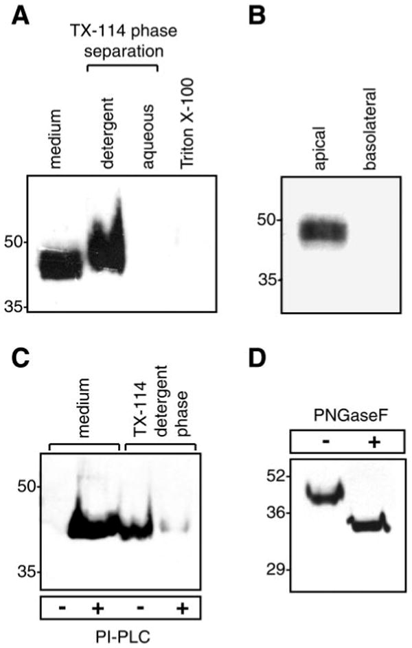
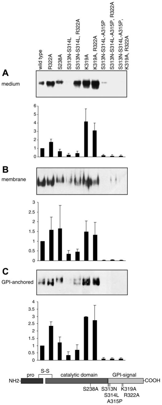
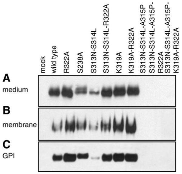
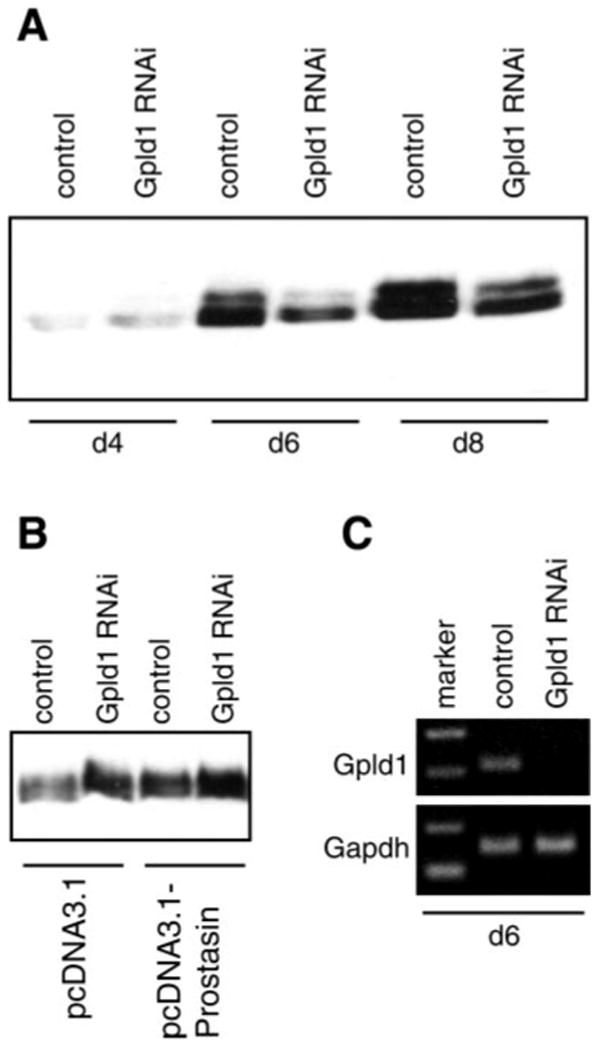
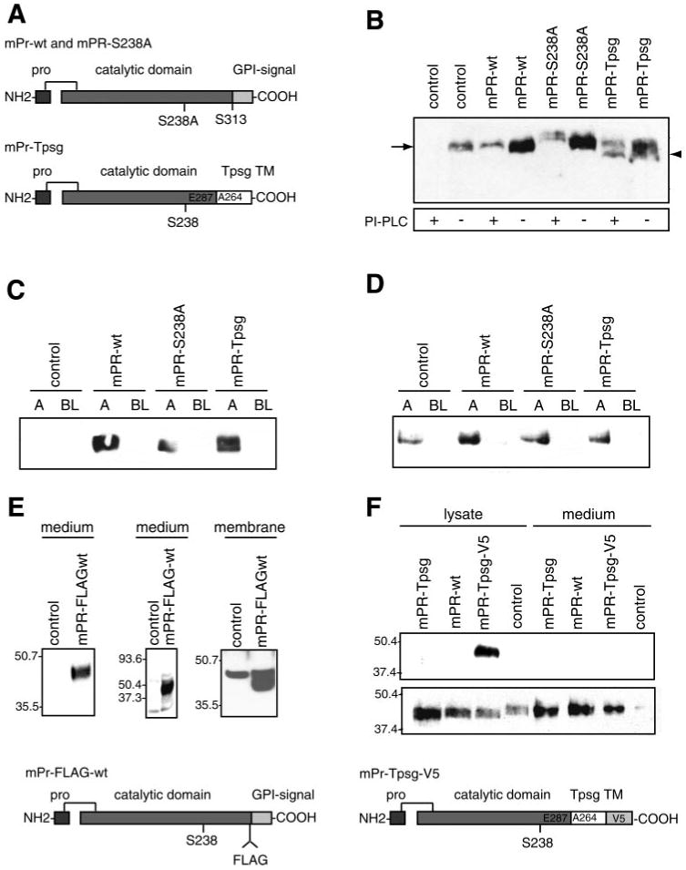
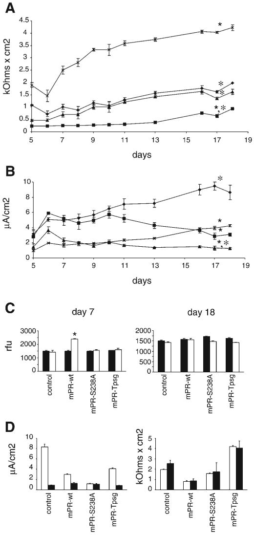
Prostasin, a trypsinlike serine peptidase, is highly expressed in prostate, kidney, and lung epithelia, where it is bound to the cell surface, secreted, or both. Prostasin activates the epithelial sodium channel (ENaC) and suppresses invasion of prostate and breast cancer cells. The studies reported here establish mechanisms of membrane anchoring and secretion in kidney and lung epithelial cells and demonstrate a critical role for prostasin in regulating epithelial monolayer function. We report that endogenous mouse prostasin is glycosylphosphatidylinositol (GPI) anchored to the cell surface and is constitutively secreted from the apical surface of kidney cortical collecting duct cells. Using site-directed mutagenesis, detergent phase separation, and RNA interference approaches, we show that prostasin secretion depends on GPI anchor cleavage by endogenous GPI-specific phospholipase D1 (Gpld1). Secretion of prostasin by kidney and lung epithelial cells, in contrast to prostate epithelium, does not depend on COOH-terminal processing at conserved Arg(322). Using stably transfected M-1 cells expressing wild-type, catalytically inactive, or chimeric transmembrane (not GPI)-anchored prostasins we establish that prostasin regulates transepithelial resistance, current, and paracellular permeability by GPI anchor- and protease activity-dependent mechanisms. These studies demonstrate a novel role for prostasin in regulating epithelial monolayer resistance and permeability in kidney epithelial cells and, furthermore, show specifically that prostasin is a critical regulator of transepithelial ion transport in M-1 cells. These functions depend on the GPI anchor as well as the peptidase activity of prostasin. These studies suggest that cell-specific Gpld1- or peptidase-dependent pathways for prostasin secretion may control prostasin functions in a tissue-specific manner.


Figures

Fig. 1
Fig. 1
Endogenous expression and posttranslational modifications of prostasin in M-1 kidney epithelial cells. A: immunoblot of native prostasin. Proteins were extracted from M-1 cells in 1% Triton X-100 or by detergent phase separation using 2% Triton X-114 (TX-114). Samples were normalized for cell number and separated by SDS-PAGE. B: immunoblot for prostasin in apical and basolateral conditioned media from M-1 cells grown to confluence [transepithelial resistance (Rte) > 1,000 Ω × cm2] on 0.4-μm-pore filters. C: glycosylphosphatidylinositol (GPI) anchoring of prostasin. M-1 cells were treated with phosphatidylinositol-specific phospholipase C (PI-PLC) or PBS. Prostasin was assayed in conditioned medium and detergent phase cell lysates by immunoblotting. D: deglycosylation of prostasin. M-1 cells were treated with PI-PLC, concentrated conditioned medium was incubated with protein N-glycosidase F (PNGase F), and proteins were separated by SDS-PAGE and immunoblotted to detect prostasin.
Fig. 2
Fig. 2
Determinants of secretion and GPI anchoring of mouse prostasin in M-1 cells. Specific amino acid mutations were introduced into prostasin to disrupt residues hypothesized to regulate GPI anchoring and secretion of prostasin as shown in the schematic diagram. Samples from M-1 cells transiently transfected with prostasin mutants were analyzed by immunoblotting and densitometry. A: conditioned medium was concentrated and normalized to load equivalent volumes; results were identical when loading was normalized to cell number. B: proteins were extracted from transfected M-1 cells by Triton X-114 detergent phase separation, precipitated with 15% trichloroacetic acid (TCA), and normalized to cell number for analysis. C: transiently transfected M-1 cells were treated with PI-PLC to release cell surface GPI-anchored proteins. Conditioned medium was concentrated, and gel loading was normalized to cell number. All experiments were done at least 3 times, and representative blots are shown. Prostasin in each compartment was quantified by densitometry and data is expressed as ratio of prostasin mutation to wild-type densitometric units. Data are expressed as means ± SD (n = 3–7).
Fig. 3
Fig. 3
Determinants of secretion and GPI anchoring of prostasin in mouse lung epithelial cells. MLE-12 cells were transiently transfected with mutated prostasins, separated by SDS-PAGE, and immunoblotted for prostasin. A: conditioned medium was concentrated and normalized to load equivalent volumes; results were identical when loading was normalized to cell number. B: proteins were extracted from MLE-12 cells by Triton X-114 detergent phase separation and precipitated with 15% TCA for analysis. Samples were normalized to cell number for immunoblotting. C: MLE-12 cells were treated with PI-PLC to release cell surface GPI-anchored proteins. Conditioned medium was concentrated, and gel loading was normalized to cell number. All experiments were done at least 3 times, and representative blots are shown. Mock transfected MLE-12 cells (no plasmid) do not express prostasin.
Fig. 4
Fig. 4
GPI-specific phospholipase D (Gpld1)-dependent secretion of prostasin. M-1 cells were transfected with Gpld1-specific small interfering RNA (siRNA) or control siRNA. A: conditioned medium was collected at 48-h intervals, concentrated, normalized to cell number, and assayed for prostasin by immunoblotting. There was no difference in cell proliferation between Gpld1- and control siRNA-transfected cells (data not shown). Relative levels of prostasin in conditioned medium were quantified by densitometry and expressed as means ± SD. Transfection with Gpld1 siRNA reduced prostasin in conditioned medium by 74% (P < 0.05; n = 3). B: M-1 cells stably transfected to express prostasin or empty vector (see Experimental Procedures and Fig. 5 for details) were transfected with Gpld1 or control siRNA and treated with PI-PLC. Medium was assayed for prostasin by immunoblotting. Inhibition of Gpld1 increased PI-PLC-releasable prostasin. C: expression of Gpld1 was assessed by semiquantitative RT-PCR relative to glyceraldehyde-3-phosphate dehydrogenase (GAPDH) control 6 days after transfection with Gpld1 and control siRNA. d4, d6, d8, 4, 6, and 8 days after transfection.
Fig. 5
Fig. 5
Characterization of stably transfected M-1 cell lines. M-1 cells were stably transfected with pcDNA3.1 empty vector (control), wild-type prostasin (mPR-wt), catalytically inactive prostasin (mPR-S238A), or chimeric transmembrane (TM), non-GPI-anchored prostasin (mPR-Tpsg). Prostasin was assayed by immunoblotting. Samples were normalized to cell number for conditioned media and protein concentration for cell lysates. A: schematic diagrams of prostasin expression constructs. B: membrane association and GPI anchoring of prostasin variants. Cells were treated with PI-PLC or buffer alone, extracted in 60 mM n-octylglucoside-1% Triton X-100 in TBS on ice, resolved by SDS-PAGE, and assayed for prostasin by immunoblotting. mPR-Tpsg (arrowhead) is resistant to shedding by PI-PLC and migrates slightly faster than prostasin (arrow). C: apical (A) and basolateral (BL) secretion of prostasin variants from stably transfected, polyclonal M-1 cell lines cultured on 0.4-μm-pore filters. D: domain-selective cell surface biotinylation. M-1 cells were grown on 0.4-μm-pore Transwell filters until stable transepithelial resistance (Rte) developed. Cells were incubated with sulfo-NHS-biotin (0.5 mg/ml) or PBS control (data not shown) in apical or basolateral chamber. Biotinylated proteins were captured with streptavidin-agarose, resolved by 10% SDS-PAGE, and assayed for prostasin by immunoblotting. E: M-1 cells were transfected with pcDNA3.1-mPR-FLAG (diagram of construct at bottom of figure) or control vector. Conditioned medium was assayed for prostasin by immunoblot with anti-prostasin antibody (left), immunoprecipitation with anti-FLAG M2 antibody followed by immunoblot with prostasin antibody (middle), or immunoblot with M2 antibody after cell surface proteins were isolated by biotinylation and streptavidin-agarose affinity capture (right). F: M-1 cells were transfected with pcDNA3.1-mPR-Tpsg, pcDNA3.1-mPR-wt, pcDNA4-mPR-Tpsg-V5 (diagram of construct at bottom of figure), or control. Cell lysates and conditioned media were assayed by immunoblot using V5 antibody (top) or prostasin antibody (bottom).
Fig. 6
Fig. 6
Effect of GPI anchoring and catalytic activity of prostasin on transepithelial resistance, current, and permeability. Stably transfected M-1 cell lines (described in Results and Fig. 5) were grown on 0.4-μm polycarbonate filters. Rte and potential difference were measured with an epithelial voltohmeter, and equivalent short-circuit current (Ieq) was calculated by Ohm's law. Data are expressed as means ± SE from 5 independent experiments with triplicate wells. Data were analyzed when resistance and potential difference were stable and monolayers of all cell lines were impermeable to inulin (data in C). A: time course of Rte in stable transfected M-1 cells overexpressing prostasin variants (♦, mPR-ctrl; ■, mPR-wt; ▲, mPR-S238A; ×, mPR-Tpsg). ★P < 0.05 compared with mPR-ctrl; *P < 0.05 compared with mPR-Tpsg. B: time course of Ieq in stably transfected M-1 cell lines. ★P < 0.05 compared with mPR-ctrl; *P < 0.05 compared with mPR-Tpsg. C: paracellular permeability of M-1 cell lines at days 7 and 18 of culture was assayed by measuring apical-to-basal flux of FITC-inulin (20 μg/ml) over 24 h. Data are expressed as means ± SE relative fluorescence units (rfu) in basal medium at 0 (filled bars) and 24 (open bars) h. Mean apical and basal fluorescence at time 0 were ∼23,000 and ∼1,600 rfu, respectively. ★P < 0.05 for comparison between 0 and 24 h. D: effect of amiloride on Ieq (left) and Rte (right) in stably transfected M-1 cell lines. Ten micromolar amiloride was applied to the apical surface of cell monolayers on days 18–19 of culture. Ieq and Rte were measured 45–60 min after exposure to amiloride. Open and filled bars depict baseline and postamiloride measurements, respectively. Data are expressed as means ± SE from 5 independent experiments with triplicate wells. Exposure to amiloride abolished the differences in Ieq between cell lines and reduced Ieq to similarly low current (P > 0.05). There was no difference in baseline and postamiloride Rte in any of these cell lines (P > 0.05).

Similar articles

Cited by

References

    1. Bauvois B. Transmembrane proteases in cell growth and invasion: new contributors to angiogenesis? Oncogene. 2004;23:317–329. - PubMed
    1. Bhagwandin V, Hau L, Clair JMS, Wolters P, Caughey G. Structure and activity of human pancreasin, a novel tryptic serine peptidase expressed primarily by the pancreas. J Biol Chem. 2003;278:3363–3371. - PubMed
    1. Brown DA, Rose JK. Sorting of GPI-anchored proteins to glycolipid-enriched membrane subdomains during transport to the apical cell surface. Cell. 1992;68:533–544. - PubMed
    1. Caughey GH, Raymond WW, Blount JL, Hau LW, Pallaoro M, Wolters PJ, Verghese GM. Characterization of human gamma-tryptases, novel members of the chromosome 16p mast cell tryptase and prostasin gene families. J Immunol. 2000;164:6566–6575. - PubMed
    1. Chen LM, Chai KX. Prostasin serine protease inhibits breast cancer invasiveness and is transcriptionally regulated by promoter DNA methylation. Int J Cancer. 2002;97:323–329. - PubMed

Publication types