. 2006 Jul 5;26(27):7257-64.
doi: 10.1523/JNEUROSCI.0196-06.2006.

BH3-only proapoptotic Bcl-2 family members Noxa and Puma mediate neural precursor cell death

Affiliations

BH3-only proapoptotic Bcl-2 family members Noxa and Puma mediate neural precursor cell death

Rizwan S Akhtar et al. J Neurosci. .

Abstract

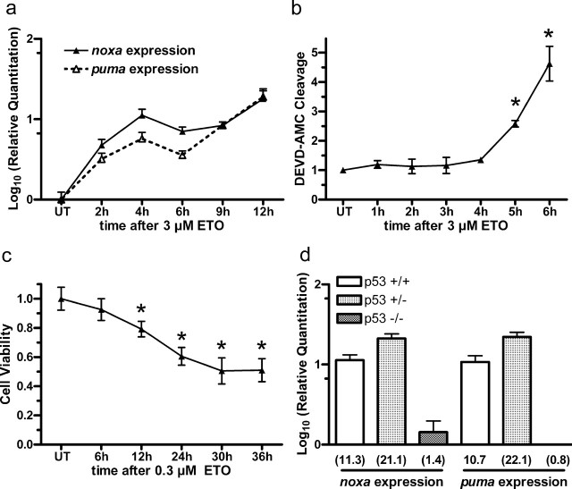
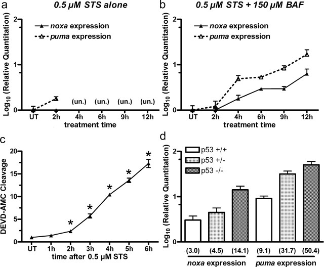
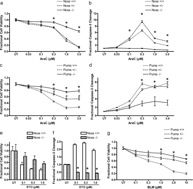
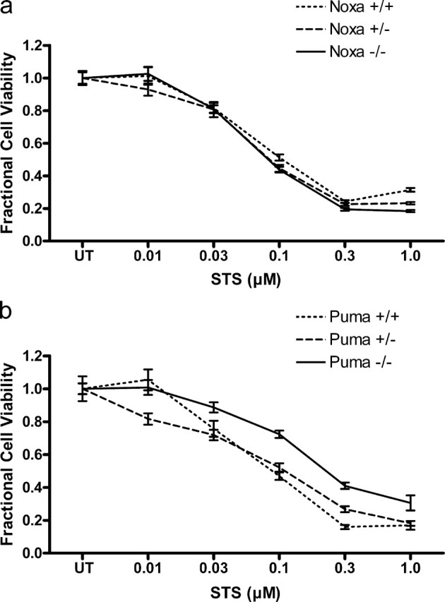
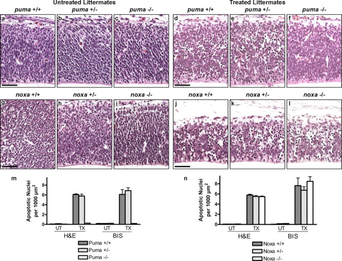
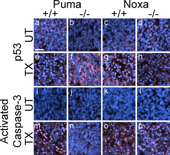
Neural precursor cells (NPCs) are highly sensitive to genotoxic injury, which triggers activation of the intrinsic mitochondria-dependent apoptotic pathway. This pathway is typically initiated by members of the BH3 (Bcl-2 homology 3)-only subgroup of the Bcl-2 (B-cell CLL/lymphoma 2) protein family, which are positioned upstream in the apoptotic pathway to respond to specific death stimuli. We have shown previously that NPCs deficient in the tumor suppressor protein p53 show significantly less death after exposure to genotoxic injury or to staurosporine (STS), a broad kinase inhibitor and potent apoptosis inducer. p53 has been shown to regulate the expression of both Noxa and Puma, two BH3-only proteins, although their involvement in p53-dependent cell death appears to be cell-type and stimulus specific. A systematic comparison of the relative contributions of Noxa and Puma to NPC apoptosis has not yet been performed. We hypothesized that p53-dependent transcription of Noxa and Puma leads to death in telencephalic NPCs exposed to genotoxic stress. We found that genotoxic injury induces a rapid p53-dependent increase in expression of Noxa and Puma mRNA in telencephalic NPCs. Furthermore, deficiency of either Noxa or Puma inhibited DNA damage-induced caspase-3 activation and cell death in telencephalic NPCs in vitro. However, only Puma deficiency protected telencephalic ventricular zone NPCs from death in vivo. In contrast to genotoxic injury, STS produced a p53-independent increase in Noxa and Puma expression, but neither Noxa nor Puma was required for STS-induced NPC death. Together, these experiments identify Noxa and Puma as important regulators of genotoxin-induced telencephalic NPC death.


Figures

Figure 1.
Figure 1.
Genotoxic stimuli induce Noxa and Puma mRNA expression in NPCs through a p53-dependent mechanism. a, An increase in Noxa mRNA expression was detected within 2 h of exposure to 3 μm ETO. Similarly, an increase in Puma mRNA expression was detected within 2 h of exposure to 3 μm ETO. b, DEVD-AMC cleavage did not significantly increase until 5–6 h after exposure to 3 μm ETO. c, Cell viability decreased significantly only after 12–36 h exposure to 0.3 μm ETO. d, After 6 h of exposure to 3 μm AraC, Noxa and Puma expression increased in wild-type and p53-heterozygous NPCs but not in p53-deficient NPCs. Experiments in a and d were performed in the presence of 150 μm BAF, a broad-spectrum caspase inhibitor, to inhibit apoptosis and thereby maximize recovery of mRNA. Levels of mRNA expression are shown relative to untreated controls (UT) and are indicated in parentheses below each column. Transcripts for either Noxa or Puma are normalized to 18S expression within each sample. No Noxa or Puma transcripts were detectable by real time RT-PCR in Noxa-deficient or Puma-deficient NPCs, respectively (data not shown). Data points in b and c represent mean ± SEM, with n = 8. ∗p < 0.001 by one-way ANOVA/Bonferroni's post hoc test versus untreated controls.
Figure 2.
Figure 2.
STS induces Noxa and Puma expression in NPCs independently of p53. a, Exposure to 0.5 μm STS did not significantly increase Noxa or Puma mRNA expression at 2 h. By 4 h, exposure to 0.5 μm STS produced alterations in 18S expression, and Noxa and Puma mRNA induction was un-interpretable (un.). b, In the presence of 150 μm BAF, modest Noxa and Puma mRNA expression was seen by 4–6 h of exposure to 0.5 μm STS. c, DEVD-AMC cleavage significantly increased within 2–3 h after exposure to 0.5 μm STS. d, After treatment with 0.5 μm STS for 6 h, Noxa and Puma mRNA expression was induced in wild-type, p53-heterozygous, and p53-deficient NPCs. Experiments in b–d were done in the presence of 150 μm BAF. UT, Untreated controls.
Figure 3.
Figure 3.
Loss of Noxa or Puma attenuates DNA damage-induced NPC death in vitro. a, NPCs that are deficient in Noxa underwent significantly less death after 36 h of exposure to AraC than wild-type or Noxa-heterozygous (Noxa+/−) NPCs. b, Induction of caspase-3-like enzymatic activity was dramatically reduced in Noxa-deficient NPCs exposed to 36 h of AraC compared with wild-type NPCs. c, Deficiency of Puma significantly protected NPCs from 24 h AraC-induced cell death. An intermediate protective effect was seen in Puma-heterozygous (Puma+/−) NPCs. d, Caspase-3-like activity was significantly attenuated by Puma deficiency in NPCs exposed to 24 h AraC. e, Loss of Noxa also protected NPCs from death after exposure to 36 h ETO. f, Compared with wild-type NPCs, caspase-3-like activity was reduced in NPCs that are deficient in Noxa after exposure to 36 h ETO. g, Loss of Puma protected NPCs from 36 h BLM-induced death. Cell viability and caspase-3-like activity were normalized to untreated controls (UT). ∗p < 0.001 by two-way ANOVA/Bonferroni's post hoc test compared with wild-type treated group. #p < 0.001 by two-way ANOVA/Bonferroni's post hoc test compared with both the wild-type and the knock-out treated group.
Figure 4.
Figure 4.
Noxa and Puma are not essential for STS-induced NPC death. a, Exposure to STS for 6 h induced significant cell death in wild-type NPCs, and this death was not affected by loss of Noxa. b, Similarly, loss of Puma did not significantly affect death after 6 h of exposure to STS. Cell viability was normalized to untreated controls (UT).
Figure 5.
Figure 5.
Puma is required for DNA damage-induced telencephalic NPC death in vivo. a–c, Untreated wild-type, Puma-heterozygous (Puma+/−), or Puma-deficient (Puma−/−) E13 embryos show scarce apoptotic cells in the ventricular zone. d, e, Wild-type or Puma+/− E13 embryos exposed to 25 mg/kg AraC for 6 h in utero exhibited widespread apoptotic cellular features in the ventricular zone. f, In contrast, littermate embryos that are deficient for Puma displayed reduced numbers of apoptotic NPCs. Apoptotic NPCs are indicated with arrows. g–i, Untreated wild-type, Noxa-heterozygous (Noxa+/−), or Noxa-deficient (Noxa−/−) E12 embryos have few apoptotic ventricular zone cells. j, k, Wild-type or Noxa+/− E12 embryos treated as above exhibited widespread apoptotic features in the ventricular zone. l, In contrast to Puma deficiency, Noxa deficiency did not significantly rescue NPCs from genotoxin-induced apoptosis. m, n, Apoptotic cells in both hematoxylin and eosin-stained (H&E) and bisbenzimide-stained (BIS) sections were quantitated in four randomly selected fields taken from multiple (n = 2) animals per genotype and expressed relative to ventricular zone area. Scale bars, 50 μm. UT, Untreated controls; TX, treated animals.
Figure 6.
Figure 6.
Loss of Puma affects p53 immunoreactivity and caspase-3 activation in telencephalic NPCs after genotoxic injury. a–d, Untreated (UT) wild-type, Puma-deficient (Puma−/−), or Noxa-deficient (Noxa−/−) embryos had low levels of p53 immunoreactivity. i–l, Untreated embryos also exhibited low immunoreactivity for activated caspase-3. e, g, m, o, Wild-type E13 embryos exposed to 25 mg/kg AraC for 6 h in utero (TX) exhibited detectable p53 immunoreactivity (e, g) and widespread immunoreactivity for activated caspase-3 (m, o) in the ventricular zone. In comparison, AraC-treated Puma-deficient littermate embryos displayed increased p53 immunoreactivity (f) and reduced caspase-3 activation (n). In contrast, AraC-treated Noxa-deficient embryos did not exhibit increased p53 immunoreactivity (h) and had significant activation of caspase-3 (p). All genotype comparisons were made between littermate embryos. Scale bar, 20 μm.

Similar articles

Cited by

References

    1. Akhtar RS, Roth KA (2006). Regulation of neural stem cell death. In: Neural development and stem cells (Rao MS, ed.) pp. 97–122. Totowa, NJ: Humana.
    1. Akhtar RS, Ness JM, Roth KA (2004). Bcl-2 family regulation of neuronal development and neurodegeneration. Biochim Biophys Acta 1644:189–203. - PubMed
    1. Akhtar RS, Geng Y, Klocke BJ, Roth KA (2006). Neural precursor cells possess multiple p53-dependent apoptotic pathways. Cell Death Differ in press. - PubMed
    1. Bounhar Y, Tounekti O, LeBlanc AC (2002). Monitoring caspases in neuronal cell death. In: Apoptosis techniques and protocols (LeBlanc AC, ed.) pp. 35–59. Totowa, NJ: Humana.
    1. Burger RM (1998). Cleavage of nucleic acids by bleomycin. Chem Rev 98:1153–1169. - PubMed

Publication types

MeSH terms