

Dopamine D3 receptor agonist delivery to a model of Parkinson's disease restores the nigrostriatal pathway and improves locomotor behavior
- PMID: 16822985
- PMCID: PMC6673939
- DOI: 10.1523/JNEUROSCI.0837-06.2006
Dopamine D3 receptor agonist delivery to a model of Parkinson's disease restores the nigrostriatal pathway and improves locomotor behavior
Abstract
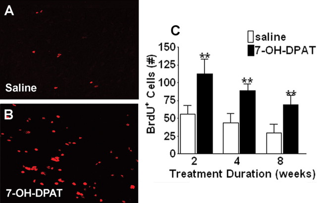
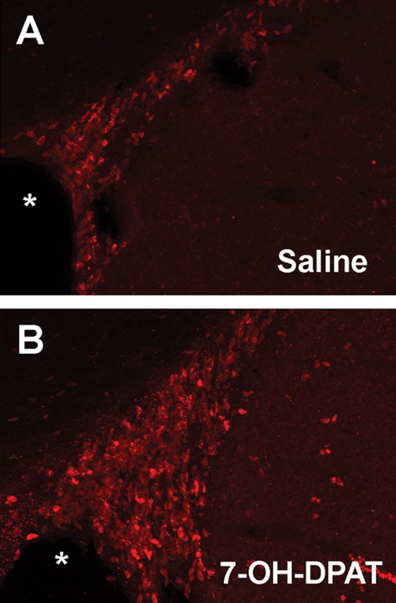
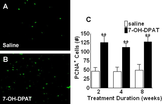
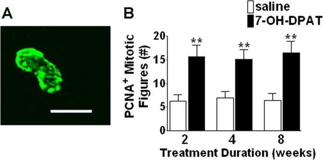
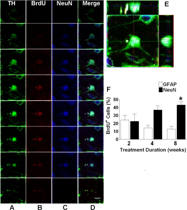
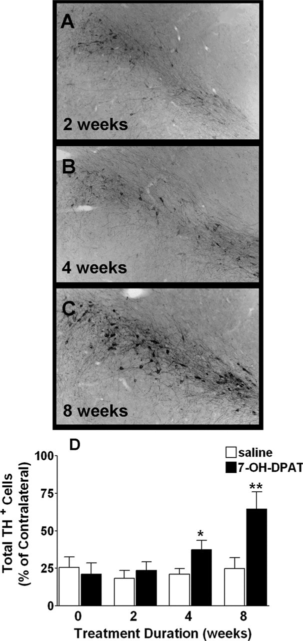
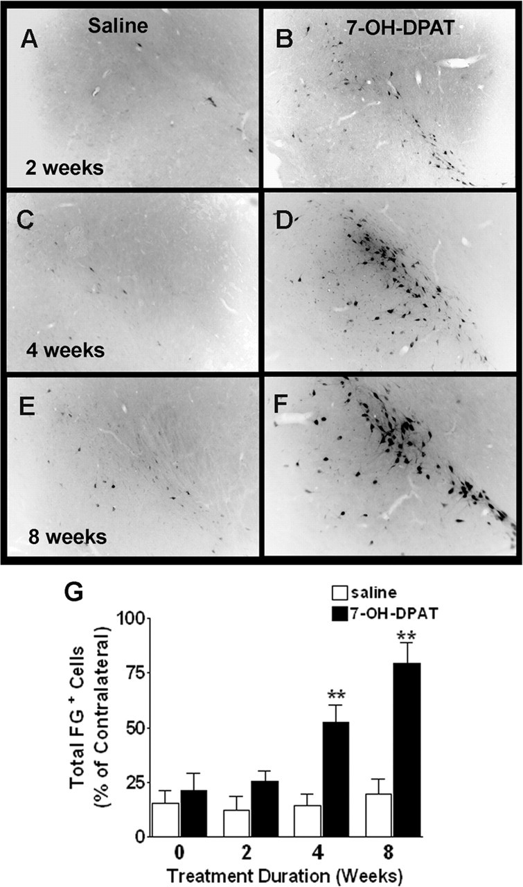
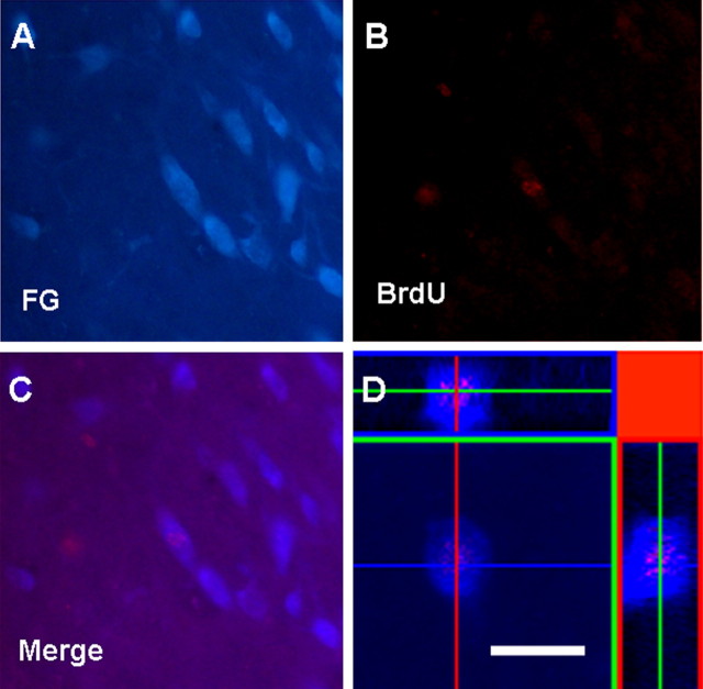
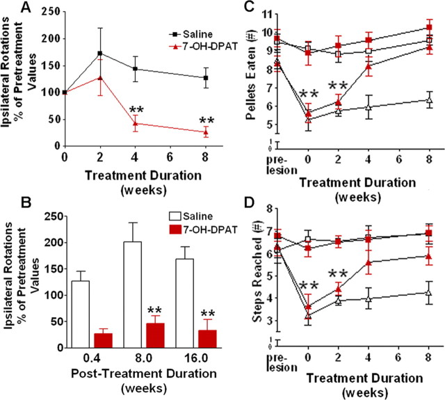
The presence of endogenous stem cell populations in the adult mammalian CNS suggests an innate potential for regeneration and represents a potential resource for neuroregenerative therapy aimed at the treatment of neurodegenerative disorders, such as Parkinson's disease. However, it is first necessary to examine the microenvironmental signals required to activate these innate reparative mechanisms. The small molecule neurotransmitter dopamine has been shown to regulate cell cycle in developing and adult brain, and the D3 receptor is known to play an important role in dopaminergic development. Pharmacological activation of the dopamine D3 receptor has been shown to trigger neurogenesis in the substantia nigra of the adult rat brain. Here, we examined the cell proliferative, neurogenic, and behavioral effects of the dopamine D3 receptor agonist 7-OH-DPAT (7-hydroxy-N,N-di-n-propyl-2-aminotetralin) in a 6-hydroxydopamine model of Parkinson's disease. Consistent with previous findings, we observed a significant induction of cell proliferation in the substantia nigra pars compacta (SN(C)) with a time-dependent adoption of a neuronal dopaminergic phenotype in many of these cells. Indices of nigrostriatal integrity were also affected. Dopaminergic cell counts in the lesioned SN(C) recovered substantially in a time-dependent manner. Similarly, retrograde tracing revealed a restoration of striatal innervation from these cells, with evidence for projections arising from newly generated cells. Finally, we observed a substantial and persistent recovery of locomotor function in these animals. The results of these studies will further our understanding of the environmental signals regulating neurogenesis in the adult brain and could have significant implications for the design of novel treatment strategies for Parkinson's disease.
Figures
Similar articles
-
A possible role for dopamine D3 receptor stimulation in the induction of neurogenesis in the adult rat substantia nigra.Neuroscience. 2005;136(2):381-6. doi: 10.1016/j.neuroscience.2005.07.054. Epub 2005 Oct 10. Neuroscience. 2005. PMID: 16216425
-
The transfection of BDNF to dopamine neurons potentiates the effect of dopamine D3 receptor agonist recovering the striatal innervation, dendritic spines and motor behavior in an aged rat model of Parkinson's disease.PLoS One. 2015 Feb 18;10(2):e0117391. doi: 10.1371/journal.pone.0117391. eCollection 2015. PLoS One. 2015. PMID: 25693197 Free PMC article.
-
Dopamine D3 receptor-preferring agonists increase dendrite arborization of mesencephalic dopaminergic neurons via extracellular signal-regulated kinase phosphorylation.Eur J Neurosci. 2008 Oct;28(7):1231-40. doi: 10.1111/j.1460-9568.2008.06423.x. Eur J Neurosci. 2008. PMID: 18973551
-
Dopamine D3 receptor agonists for protection and repair in Parkinson's disease.Curr Opin Pharmacol. 2007 Feb;7(1):100-5. doi: 10.1016/j.coph.2006.11.004. Epub 2006 Dec 13. Curr Opin Pharmacol. 2007. PMID: 17174156 Review.
-
Dopamine D3 agonists in the treatment of Parkinson's disease.Curr Top Med Chem. 2015;15(10):908-26. doi: 10.2174/156802661510150328223428. Curr Top Med Chem. 2015. PMID: 25832718 Review.
Cited by
-
Dopaminergic agonists: possible neurorescue drugs endowed with independent and synergistic multisites of action.Neurochem Res. 2007 Oct;32(10):1726-9. doi: 10.1007/s11064-007-9350-9. Epub 2007 May 8. Neurochem Res. 2007. PMID: 17486445 Review.
-
Restoration of nigrostriatal dopamine neurons in post-MPTP treatment by the novel multifunctional brain-permeable iron chelator-monoamine oxidase inhibitor drug, M30.Neurotox Res. 2010 Jan;17(1):15-27. doi: 10.1007/s12640-009-9070-9. Epub 2009 Jul 16. Neurotox Res. 2010. PMID: 19609632
-
Role of Dopamine D2/D3 Receptors in Development, Plasticity, and Neuroprotection in Human iPSC-Derived Midbrain Dopaminergic Neurons.Mol Neurobiol. 2018 Feb;55(2):1054-1067. doi: 10.1007/s12035-016-0376-3. Epub 2017 Jan 14. Mol Neurobiol. 2018. PMID: 28092083
-
Adult Endogenous Dopaminergic Neuroregeneration Against Parkinson's Disease: Ideal Animal Models?Neurotox Res. 2021 Apr;39(2):504-532. doi: 10.1007/s12640-020-00298-7. Epub 2020 Nov 3. Neurotox Res. 2021. PMID: 33141428 Review.
-
Effect of cyclosporin A on the uptake of D3-selective PET radiotracers in rat brain.Nucl Med Biol. 2011 Jul;38(5):725-39. doi: 10.1016/j.nucmedbio.2011.01.002. Epub 2011 Mar 3. Nucl Med Biol. 2011. PMID: 21718948 Free PMC article.
References
-
- Abrous DN, Dunnett SB (1994). Skilled paw reaching in rats: the staircase test. Neurosci Protoc 3:1–11.
-
- Baker SA, Baker KA, Hagg T (2004). Dopaminergic nigrostriatal projections regulate neural precursor cell proliferation in the adult mouse subventricular zone. Eur J Neurosci 20:575–579. - PubMed
-
- Barnéoud P, Parmentier S, Mazadier M, Miquet JM, Boireau A, Dubédat P, Blanchard JC (1995). Effects of complete and partial lesions of the dopaminergic mesotelencephalic system on skilled forelimb use in the rat. Neuroscience 67:837–848. - PubMed
-
- Bartlett LE, Mendez I (2005). Dopaminergic reinnervation of the globus pallidus by fetal nigral grafts in the rodent model of Parkinson's disease. Cell Transplant 14:119–127. - PubMed
-
- Bjorklund A, Sanchez-Pernaute R, Chung S, Andersson T, Chen IY, McNaught KS, Brownell AL, Jenkins BG, Wahlstedt C, Kim KS, Isacson O (2002). Embryonic stem cells develop into functional dopaminergic neurons after transplantation in a Parkinson's rat model. Proc Natl Acad Sci USA 99:2344–2349. - PMC - PubMed
Publication types
MeSH terms
Substances
LinkOut - more resources
Full Text Sources
Other Literature Sources
Miscellaneous