Comparative Study
. 2006 Aug;15(8):1842-8.
doi: 10.1110/ps.062278006. Epub 2006 Jul 5.

Structure of the angiogenesis inhibitor ovalicin bound to its noncognate target, human Type 1 methionine aminopeptidase

Affiliations
Comparative Study

Structure of the angiogenesis inhibitor ovalicin bound to its noncognate target, human Type 1 methionine aminopeptidase

Anthony Addlagatta et al. Protein Sci. 2006 Aug.

Abstract

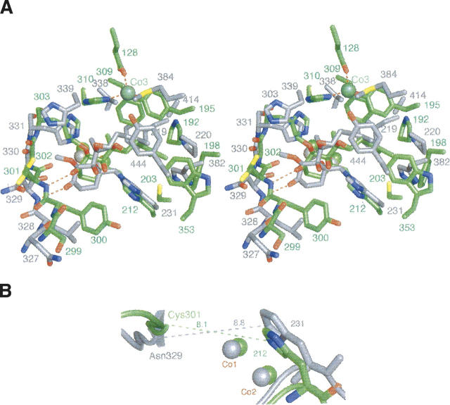
Methionine aminopeptidases (MetAPs) remove the initiator methionine during protein biosynthesis. They exist in two isoforms, MetAP1 and MetAP2. The anti-angiogenic compound fumagillin binds tightly to the Type 2 MetAPs but only weakly to Type 1. High-affinity complexes of fumagillin and its relative ovalicin with Type 2 human MetAP have been reported. Here we describe the crystallographic structure of the low-affinity complex between ovalicin and Type 1 human MetAP at 1.1 A resolution. This provides the first opportunity to compare the structures of ovalicin or fumagillin bound to a Type 1 and a Type 2 MetAP. For both Type 1 and Type 2 human MetAPs the inhibitor makes a covalent adduct with a corresponding histidine. At the same time there are significant differences in the alignment of the inhibitors within the respective active sites. It has been argued that the lower affinity of ovalicin and fumagillin for the Type 1 MetAPs is due to the smaller size of their active sites relative to the Type 2 enzymes. Comparison with the uncomplexed structure of human Type 1 MetAP indicates that there is some truth to this. Several active site residues have to move "outward" by 0.5 Angstroms or so to accommodate the inhibitor. Other residues move "inward." There are, however, other factors that come into play. In particular, the side chain of His310 rotates by 134 degrees into a different position where (together with Glu128 and Tyr195) it coordinates a metal ion not seen at this site in the native enzyme.


Figures

Figure 1.
Figure 1.
Structures of the natural products fumagillin and ovalicin and the synthetic derivative TNP-470.
Figure 2.
Figure 2.
(A) A stereo view of an “omit” electron density map within the active site of tHsMetAP1-ov. Coefficients are (Fo–Fc), where Fo are observed amplitudes. The calculated amplitudes Fc and phases were obtained from the refined model with ovalicin removed. The map is calculated at 1.1 Å resolution and contoured at 4.2 σ. The spiroepoxy group of ovalicin covalently modifies His212. The isoprenyl group is buried deep in the active site and surrounded by several hydrophobic residues. In the ribbon diagram, the catalytic domain of the protein is depicted in gray; the N-terminal region, in red. (B) Ball-and-stick representation of ovalicin (yellow) covalently linked to His212 of tHsMetAP1. The newly released hydroxyl group forms a hydrogen bond with the bridging water/hydroxo anion (red) between the active site cobalt ions (purple). (C) ORTEP diagram of ovalicin (yellow) and His212 (green and blue). The size of the ellipsoids represents the thermal motion. The isoprenyl side chain is less ordered than the ring moiety. (D) Stereo view of the superimposed active site residues of tHsMetAP1 in the native (gray) and ovalicin-bound forms (red). Ovalicin is represented with thicker bonds. His310 rotates away from the active site providing space for the inhibitor. Similarly, several other residues surrounding the ovalicin in tHsMetAP1-ov undergo outward movement. Cys301, Phe309, and Met338 move inward.
Figure 3.
Figure 3.
Stereo diagram of the active site metal center is shown with an “omit” electron density map around Asp229. The map is contoured at 4.5 σ. The “native” conformation of Asp229 is shown in green. An alternative conformation shown in yellow is estimated to occur at 25% occupancy in the inhibitor complex.
Figure 4.
Figure 4.
(A) Stereo view of the superimposed active sites of tHsMetAP1-ov (green) and HsMetAP2-ov (gray). The relative orientation of ovalicin differs somewhat in the two structures. Also, His339 in HsMetAP2-ov moves much farther than His310 of tHsMetAP1-ov. (B) The distance between Nɛ2 of His212 and the entrance of the active site is shorter (8.1 Å) in tHsMetAP1 than that in HsMetAP2 (8.8 Å). This is the region occupied by the cyclohexanone ring of the inhibitor.
Figure 5.
Figure 5.
Superposition of the ovalicin complex with Type 2 human MetAP (gray) and the ovalicin complex with Type 1 human enzyme (green). The figure shows the vicinity of Thr334, and the numbering is for HsMetAP1-ov. In HsMetAP2-ov the hydrogen bond between the backbone of Ala362 and the backbone of His321 is shown in red. The analogous hydrogen bond in tHsMetAP1-ov between Oγ1 of Thr334 and the backbone amide of His303 is shown in green.

Similar articles

Cited by

References

    1. Addlagatta A., Hu X., Liu J.O., Matthews B.W. 2005. Structural basis for the functional differences between Type I and Type II human methionine aminopeptidases. Biochemistry 44: 14741–14749. - PubMed
    1. Bergers G., Javaherian K., Lo K.M., Folkman J., Hanahan D. 1999. Effects of angiogenesis inhibitors on multistage carcinogenesis in mice. Science 284: 808–812. - PubMed
    1. Bhargava P., Marshall J.L., Rizvi N., Dahut W., Yoe J., Figuera M., Phipps K., Ong V.S., Kato A., Hawkins M.J. 1999. A Phase I and pharmacokinetic study of TNP-470 administered weekly to patients with advanced cancer. Clin. Cancer Res. 5: 1989–1995. - PubMed
    1. Brdlik C.M. and Crews C.M. 2004. A single amino acid residue defines the difference in ovalicin sensitivity between Type I and II methionine aminopeptidases. J. Biol. Chem. 279: 9475–9480. - PMC - PubMed
    1. Brünger A.T., Adams P.D., Clore G.M., DeLano W.L., Gros P., Grosse-Kunstleve R.W., Jiang J.S., Kuszewski J., Nilges M., Pannu N.S. et al. 1998. Crystallography & NMR system: A new software suite for macromolecular structure determination. Acta Crystallogr. D Biol. Crystallogr. 54: 905–921. - PubMed

Publication types

LinkOut - more resources