

Comparative study of Domoic Acid and Okadaic Acid induced-chromosomal abnormalities in the Caco-2 cell line
- PMID: 16823071
- PMCID: PMC3785674
- DOI: 10.3390/ijerph2006030001
Comparative study of Domoic Acid and Okadaic Acid induced-chromosomal abnormalities in the Caco-2 cell line
Abstract
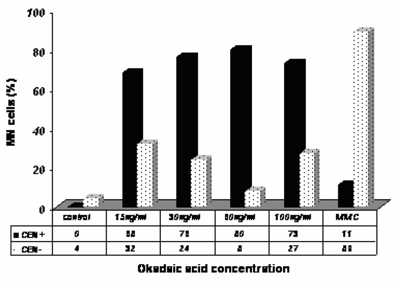
Okadaic Acid (OA) the major diarrheic shellfish poisoning (DSP) toxin is known as a tumor promoter and seems likely implicated in the genesis of digestive cancer. Little is known regarding genotoxicity and carcinogenicity of Domoic Acid (DA), the major Amnesic Shellfish Poisoning (ASP) toxin. Both OA and DA occur in seafood and are of human health concerns. Micronuclei (MN) arise from abnormalities in nuclear division during mitosis due to a failure of the mitotic spindle or by complex chromosomal configurations that pose problems during anaphase. In order to evaluate the ability of okadaic acid (OA) and domoic acid (DA) to induce DNA damage we performed the micronucleus assay using the Caco-2 cell line. To discriminate between a clastogenic or aneugenic effect of OA and DA, the micronucleus assay was conducted by cytokinesis-block micronucleus assay using cytochalasin B with Giemsa staining and/or acridine orange staining, in parallel to fluorescence in situ hybridization (FISH) using a concentrated human pan-centromeric chromosome paint probe. Our results showed that OA and DA significantly increased the frequency of MN in Caco-2 cells. The MN caused by OA are found in mononucleated cells and binucleated cells, whereas those caused by DA are mainly in binucleated cells. The results of FISH analysis showed that OA induced centromere-positive micronuclei and DA increased the percentage of MN without a centromeric signal. In conclusion, both OA and DA bear mutagenic potential as revealed in Caco-2 cells by induction of MN formation. Moreover, OA induced whole chromosome loss suggesting a specific aneugenic potential, whereas DA seems simply clastogenic. At present, one cannot rule out possible DNA damage of intestinal cells if concentrations studied are reached in vivo, since this may happen with concentrations of toxins just below regulatory limits in case of frequent consumption of contaminated shell fishes.
Figures


References
-
- Sournia A, Chretiennot D, Ricard M. Marine phytoplankton: how species in the world ocean? J. Plankton Res. 1991;13:1093–1099.
-
- Suganuma M, Suttajit M, Suguri H, Ojika M, Yamada K, Fujika H. Specific binding of okadaic acid, a tumor promoter in mouse skin. FEBS Lett. 1989;250:615–618. - PubMed
-
- Sueoka E, Fujiki HJ. Carcinogenesis of okadaic acid class tumor promoters derived from marine natural products. Cancer Res. Clin. Oncol. 1997;123:413–419.
-
- Matias WG, Traoré A, Creppy EE. Variations in the distribution of okadaic acid in organs and biological fluids of mice related to diarrhoeic syndrome. Hum. Exp. Toxicol. 1999;18:345–350. - PubMed
-
- Traore A, Ruiz S, Baudrimont I, Sanni A, Dano SD, Guarigues P, Narbonne JF, Creppy EE. Combined effects of okadaic acid an d cadmium on lipid peroxidation and DNA bases modifications (m5dC and 8-(OH)-dG) in Caco-2 cells. Arch. Toxicol. 2000;74:79–84. - PubMed
Publication types
MeSH terms
Substances
LinkOut - more resources
Full Text Sources
Miscellaneous