. 2006 Mar;3(1):11-22.
doi: 10.3390/ijerph2006030002.

Neuregulin 1-Beta cytoprotective role in AML 12 mouse hepatocytes exposed to pentachlorophenol

Affiliations

Neuregulin 1-Beta cytoprotective role in AML 12 mouse hepatocytes exposed to pentachlorophenol

Waneene C Dorsey et al. Int J Environ Res Public Health. 2006 Mar.

Abstract

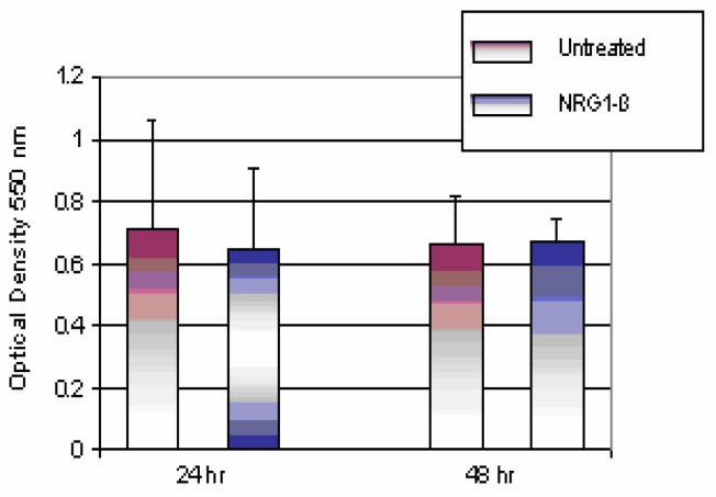
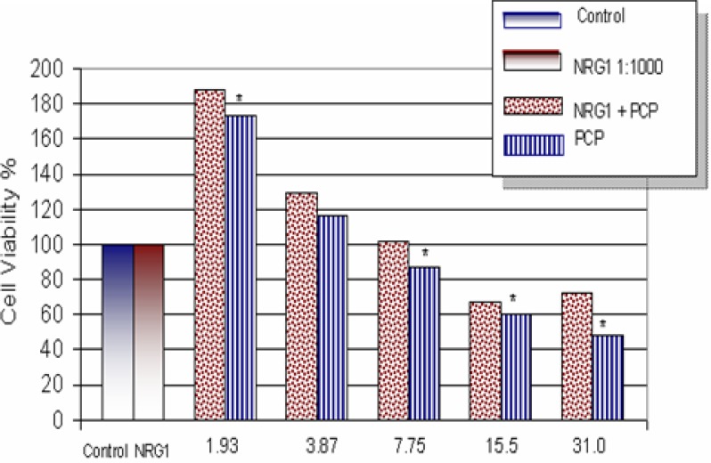
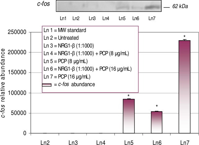
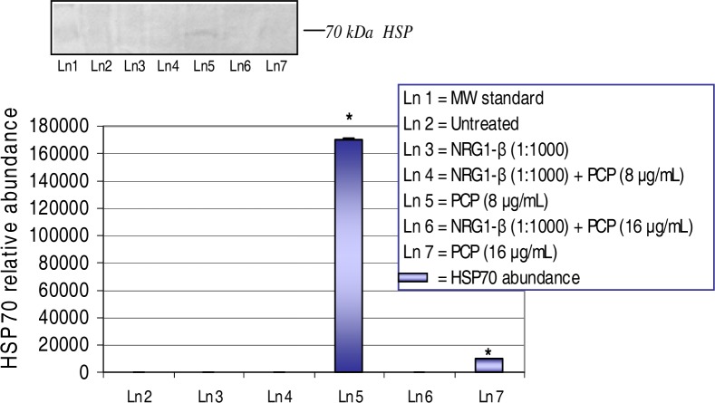
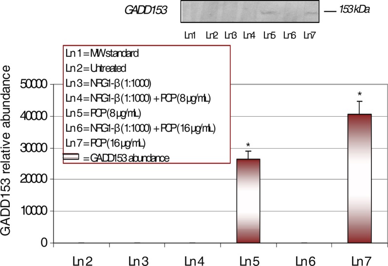
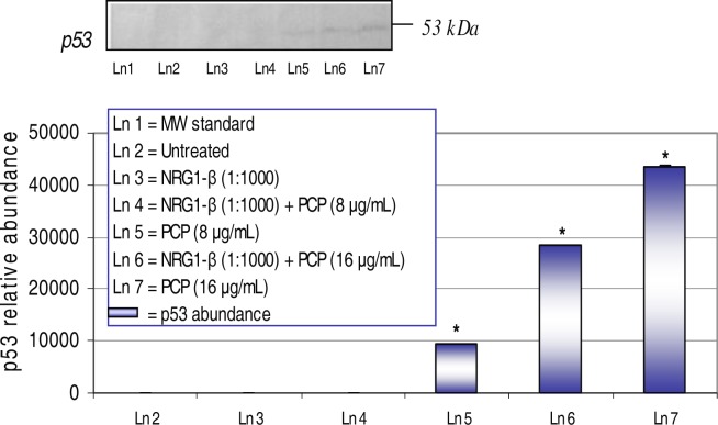
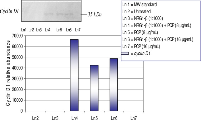
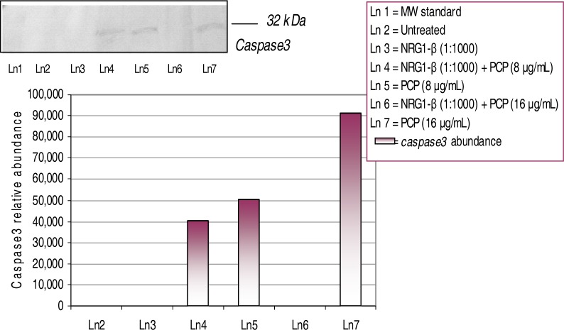
Neuregulins are a family of growth factor domain proteins that are structurally related to the epidermal growth factor. Accumulating evidence has shown that neuregulins have cyto- and neuroprotective properties in various cell types. In particular, the neuregulin-1 Beta (NRG1-Beta) isoform is well documented for its antiinflammatory properties in rat brain after acute stroke episodes. Pentachlorophenol (PCP) is an organochlorine compound that has been widely used as a biocide in several industrial, agricultural, and domestic applications. Previous investigations from our laboratory have demonstrated that PCP exerts both cytotoxic and mitogenic effects in human liver carcinoma (HepG2) cells, primary catfish hepatocytes and AML 12 mouse hepatocytes. We have also shown that in HepG2 cells, PCP has the ability to induce stress genes that may play a role in the molecular events leading to toxicity and tumorigenesis. In the present study, we hypothesize that NRG1-Beta will exert its cytoprotective effects in PCP-treated AML 12 mouse hepatocytes by its ability to suppress the toxic effects of PCP. To test this hypothesis, we performed the MTT-cell respiration assay to assess cell viability, and Western-blot analysis to assess stress-related proteins as a consequence of PCP exposure. Data obtained from 48 h-viability studies demonstrated a biphasic response; showing a dose-dependent increase in cell viability within the range of 0 to 3.87 microg/mL, and a gradual decrease within the concentration range of 7.75 to 31.0 microg/mL in concomitant treatments of NRG1-Beta+PCP and PCP. Cell viability percentages indicated that NRG1-Beta+PCPtreated cells were not significantly impaired, while PCP-treated cells were appreciably affected; suggesting that NRG1-Beta has the ability to suppress the toxic effects of PCP. Western Blot analysis demonstrated the potential of PCP to induce oxidative stress and inflammatory response (c-fos), growth arrest and DNA damage (GADD153), proteotoxic effects (HSP70), cell cycle arrest as consequence of DNA damage (p53), mitogenic response (cyclin- D1), and apoptosis (caspase-3). NRG1-Beta exposure attenuated stress-related protein expression in PCP-treated AML 12 mouse hepatocytes. Here we provide clear evidence that NRG1-Beta exerts cytoprotective effects in AML 12 mouse hepatocytes exposed to PCP.


Figures

Figure 1:
Figure 1:
Comparison of untreated and NRG1-β-treated AML 12 mouse hepatocytes. NRG1-β-treated (10 nM NRG1-β; 1:1000) hepatocytes were compared to untreated (0) cells for 24- and 48 h. Hepatocytes were maintained in DMEM medium with 10% FBS supplement. On day of exposure, FBS-medium was replaced with serum-free medium. The MTT-assay was used to determine absorbance at 550 nm after 24- and 48 h exposure periods. Absorbance readings are expressed as optical density. Each bar represents the mean ± S.D (n=3 independent experiments; p>0.05).
Figure 2:
Figure 2:
Effect of NRG1-β on PCP toxicity in AML 12 Mouse Hepatocytes. Cells were treated for 48hrs with PCP in the presence or absence of NRG1-β (0.01 nM). The number of metabolically active cells was determined by the MTT incorporation. The data are expressed as percentages of cell viability. Each point represents a mean value and standard deviation of three independent experiments (n = 3 independent experiments; 8 replications per treatment). *Significantly different (p < 0.05) from NRG1-β-treatment.
Figure 3:
Figure 3:
Expression and relative abundance of the 62 kDa c-fos in AML 12 mouse hepatocytes exposed to PCP and NRG1-β + PCP for 48 h. AML 12 mouse hepatocytes were treated with 8 µg/mL and 16 µg/mL concomitant treatments of PCP and NRG1-β + PCP. c-fos protein identification was assessed following exposure incubation period of 48 h. Inset shows a representative Wester n blot analysis. Each point represents a mean value and standard deviation of three experiments. *Significantly different (p < 0.05) from untreated (0 µg/mL PCP) and NRG1-β (1:1000) treated cells.
Figure 4:
Figure 4:
Expression and relative abundance of the 70 kDa heat shock (HSP70) in AML 12 mouse hepatocytes exposed to PCP and NRG1-β + PCP for 48 h. AML 12 mouse hepatocytes were treated with 8 μg/mL and 16 μg/mL concomitant treatments of PCP and NRG1-β+PCP. HSP70 protein identification was assessed following exposure incubation period of 48 h. Inset shows a representative Western blot analysis. Each point represents a mean value and standard deviation of three experiments. *Significantly different (p<0.05) from untreated (0 µg/mL PCP) and NRG1-β (1:1000) treated cells.
Figure 5:
Figure 5:
Expression and relative abundance of the 153 kDa GADD in AML 12 mouse hepatocytes exposed to PCP and NRG1-β + PCP for 48 h. AML 12 mouse hepatocytes were treated with 8 μg/mL and 16 μg/mL concomitant treatments of PCP and NRG1-β + PCP. GADD153 protein identification was assessed following exposure incubation period of 48 h. Inset shows representative Wester n blot analysis. Each point represents a mean value and standard deviation of three experiments. *Significantly different (p ≤ 0.05) from untreated (0 µg/mL PCP) and NRG1-β (1:1000) treated cells.
Figure 6:
Figure 6:
Expression and relative abundance of p53 in AML 12 mouse hepatocytes exposed to PCP and NRG1-β+ PCP for 48 h. p53 protein identification was assessed following exposure incubation period of 48 h. Inset shows representative Western blot analysis. Each point represents a mean value and standard deviation of three independent experiments. *Significantly different (p < 0.05) from untreated (0 μg/mL) and NRG1-β (1:1000) treated cells.
Figure 7:
Figure 7:
Expression and relative abundance of cyclin D1 in AML 12 mouse hepatocytes exposed to PCP and NRG1-β + PCP for 48h. Cyclin D1 protein identification was assessed following exposure incubation period of 48 h. Inset shows a representative Western-blot analysis. The following values were compared to untreated (0 µg/mL) and NRG1-β (1:1000) treated cells.
Figure 8:
Figure 8:
Expression and relative abundance of caspase-3 in AML 12 mouse hepatocytes exposed to PCP and NRG1-β + PCP for 48h. Caspase-3 protein identification was assessed following exposure incubation period of 48 h. Inset shows a representative Western-blot analysis. The following values were compared to untreated (0 µg/mL) and NRG1-β (1:1000) treated cells.

Similar articles

Cited by

References

    1. Crovello CS, Lai C, Cantley LC, Carraway KL., III Differential signaling by the epidermal growth factor-like growth factors neuregulin-1 and neuregulin -2. J Biol Chem. 1998;273:26954–26961. - PubMed
    1. Burden S, Yarden Y. Neuregulins and their receptors: a versatile signaling module in organogenesis and oncogenesis. Neuron. 1997;18:847–855. - PubMed
    1. Nakano N, Higashiyama S, Ohmoto H, Ishiguro H, Taniguchi N, Wada Y. The N-terminal region of NTAK/neuregulin-2 isoforms has an inhibitory activity on angiogenesis. J Biol Chem. 2004;279(12):11465–11470. - PubMed
    1. Parkinson DB, Langner K, Namini SS, Jessen KR, Mirksy R. β-Neuregulin and autocrine mediated survival of Schwann cells requires activity of Ets family transcription factors. Molecular and Cellular Neuroscience. 2002;20:154–167. - PubMed
    1. Murphy S, Krainock R, Tham M. Neuregulin signaling via ErbB receptor assemblies in the nervous system. Molecular Neurobiology. 2002;25:67–78. - PubMed