Review
. 2006 Jul;116(7):1761-6.
doi: 10.1172/JCI29063.

Central insulin action in energy and glucose homeostasis

Affiliations
Review

Central insulin action in energy and glucose homeostasis

Leona Plum et al. J Clin Invest. 2006 Jul.

Abstract

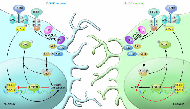
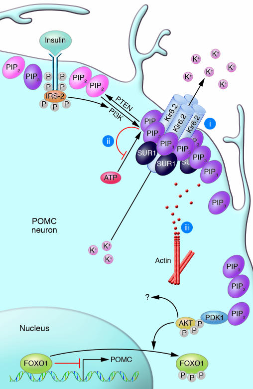
Insulin has pleiotropic biological effects in virtually all tissues. However, the relevance of insulin signaling in peripheral tissues has been studied far more extensively than its role in the brain. An evolving body of evidence indicates that in the brain, insulin is involved in multiple regulatory mechanisms including neuronal survival, learning, and memory, as well as in regulation of energy homeostasis and reproductive endocrinology. Here we review insulin's role as a central homeostatic signal with regard to energy and glucose homeostasis and discuss the mechanisms by which insulin communicates information about the body's energy status to the brain. Particular emphasis is placed on the controversial current debate about the similarities and differences between hypothalamic insulin and leptin signaling at the molecular level.


Figures

Figure 1
Figure 1. Insulin and leptin increase expression of POMC and decrease expression of AgRP.
Insulin binds to its receptor on POMC and AgRP neurons, stimulating receptor autophosphorylation and activating its signal cascade. IRS proteins bind to the phosphorylated residues on the IR and subsequently recruit the regulatory subunit p85 of PI3K. PI3K thereby becomes activated and phosphorylates phosphatidylinositol-4,5-bisphosphate (PIP2) on position 3′ in the inositol ring, generating PIP3. The lipid phosphatase PTEN antagonizes this by dephosphorylating PIP3 to generate PIP2. The protein kinase B/AKT and phosphoinositide-dependent protein kinase 1 (PDK1) bind to PIP3 via their pleckstrin homology (PH) domains. AKT is readily phosphorylated and activated by PDK1. Phosphorylated AKT enters the nucleus, where it phosphorylates FOXO1. This leads to exclusion from the nucleus and thereby to inactivation of FOXO1. FOXO1 exerts different effects in POMC and AgRP neurons. In POMC neurons, it diminishes POMC transcription by recruiting Ncor and Hdac1 and by competing with binding sites for phosphorylated STAT3 in the promoter. By phosphorylating and excluding FOXO1 from the nucleus, insulin de-inhibits the promoter, thereby increasing POMC expression. In AgRP neurons, FOXO1 increases AgRP transcription. Here insulin decreases FOXO1-mediated transcription of AgRP by excluding FOXO1 from the nucleus. Leptin binds to its receptor, leading to recruitment of JAKs, which phosphorylate the receptor. The STAT3 monomer binds to the activated leptin receptor and is phosphorylated by JAKs. Upon phosphorylation, two STATs homodimerize and translocate to the nucleus, where they activate POMC transcription in POMC neurons and decrease AgRP transcription in AgRP neurons. It is possible that leptin also induces activation of PI3K. Consequently, insulin de-inhibits and leptin activates the POMC promoter. Insulin deactivates and leptin inhibits the AgRP promoter.
Figure 2
Figure 2. Generation of PIP3 leads to KATP channel opening and consecutive cell hyperpolarization.
Insulin activates PI3K, which phosphorylates PIP2 on position 3′ in the inositol ring, generating PIP3. The lipid phosphatase PTEN antagonizes this by dephosphorylating PIP3 to generate PIP2. PIP3 accumulation leads to activation of KATP channels and, thus, to potassium outflow. This leads to membrane hyperpolarization and silencing of the neuron. Three different mechanisms for channel opening have been suggested: (i) PIP3 binding to the Kir6.2 subunit of the potassium channel increases the probability that the channel is open, which indirectly lowers inhibition by ATP; (ii) PIP3 competes with ATP for binding to the Kir6.2 subunit, thereby lowering ATP’s ability to close the channel; and (iii) PIP3 activates degradation of the local actin cytoskeleton. Also, activation of proteins downstream in the insulin cascade such as PDK1, AKT, glycogen synthase kinase 3 (GSK3), or mammalian target of rapamycin (mTOR) may be involved in insulin’s ability to regulate KATP channel opening.

References

    1. Bernard C.1855. . Leçons de physiologie expérimentale appliquée à la médecine faites au Collège de France. Baillère et Fils.Paris, France.296–313.
    1. Rulifson E.J., Kim S.K., Nusse R. Ablation of insulin-producing neurons in flies: growth and diabetic phenotypes. Science. 2002;296:1118–1120. - PubMed
    1. Wolkow C.A., Kimura K.D., Lee M.S., Ruvkun G. Regulation of C. elegans life-span by insulinlike signaling in the nervous system. Science. 2000;290:147–150. - PubMed
    1. Cheng C.L., Gao T.Q., Wang Z., Li D.D. Role of insulin/insulin-like growth factor 1 signaling pathway in longevity. World J. Gastroenterol. 2005;11:1891–1895. - PMC - PubMed
    1. Tissenbaum H.A., Ruvkun G. An insulin-like signaling pathway affects both longevity and reproduction in Caenorhabditis elegans. Genetics. 1998;148:703–717. - PMC - PubMed

Publication types