

Neuropeptide signaling and hydrocephalus: SCO with the flow
- PMID: 16823482
- PMCID: PMC1483144
- DOI: 10.1172/JCI29148
Neuropeptide signaling and hydrocephalus: SCO with the flow
Abstract
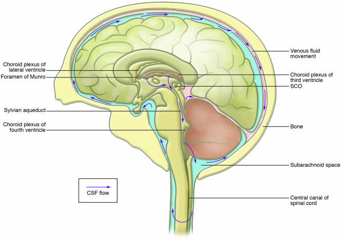
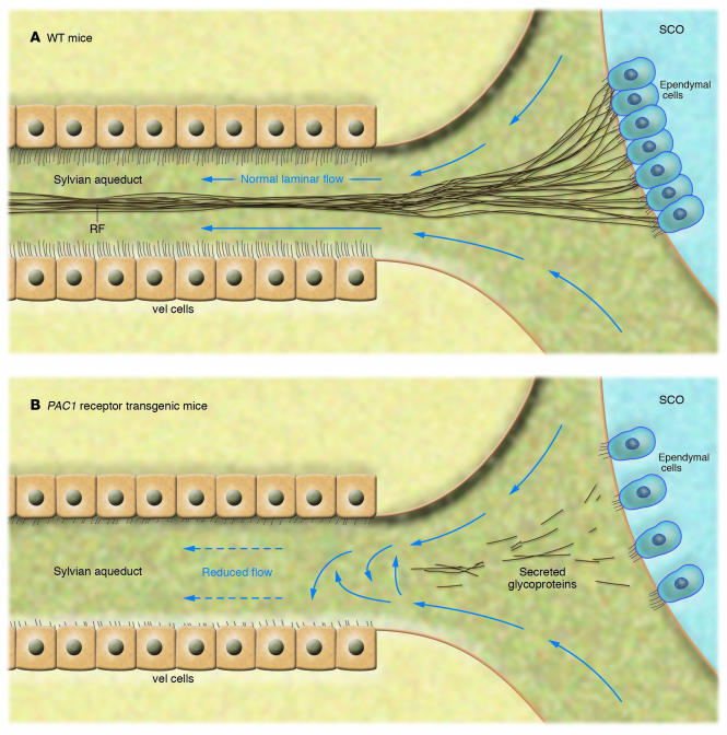
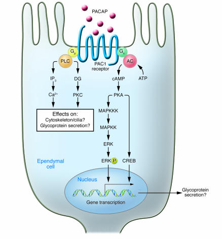
Congenital hydrocephalus affects 0.1-0.3% of live births, with a high mortality rate (approximately 50%) in the absence of surgical intervention. Although the insertion of shunts alleviates the symptoms of the majority of congenital cases, the molecular basis of hydrocephalus and the mechanisms of cerebrospinal fluid (CSF) circulation remain largely unknown. Two important players are the subcommissural organ/Reissner's fiber (SCO/RF) complex and the ventricular ependymal (vel) cells that together facilitate the flow of the CSF through the narrow canals of the ventricular system. In this issue of the JCI, Lang et al. demonstrate that overexpression of the pituitary adenylate cyclase-activating polypeptide (PACAP) type I (PAC1) receptor gene results in abnormal development of the SCO and vel cells, leading to congenital hydrocephalus (see the related article beginning on page 1924). The ligand for the PAC1 receptor is the neuropeptide PACAP, which uncovers what the authors believe to be a novel role for this signaling cascade in the regulation of CSF circulation.
Figures
Comment on
-
Expression of the human PAC1 receptor leads to dose-dependent hydrocephalus-related abnormalities in mice.J Clin Invest. 2006 Jul;116(7):1924-34. doi: 10.1172/JCI27597. J Clin Invest. 2006. PMID: 16823490 Free PMC article.
Similar articles
-
Subcommissural organ, cerebrospinal fluid circulation, and hydrocephalus.Microsc Res Tech. 2001 Mar 1;52(5):591-607. doi: 10.1002/1097-0029(20010301)52:5<591::AID-JEMT1043>3.0.CO;2-7. Microsc Res Tech. 2001. PMID: 11241868 Review.
-
Pituitary adenylyl cyclase-activating polypeptide (PACAP) and its receptor (PAC1-R) in the cochlea: evidence for specific transcript expression of PAC1-R splice variants in rat microdissected cochlear subfractions.Neuroscience. 2006 Jun 19;140(1):147-61. doi: 10.1016/j.neuroscience.2006.01.019. Epub 2006 Apr 19. Neuroscience. 2006. PMID: 16626868
-
SCO-ping out the mechanisms underlying the etiology of hydrocephalus.Physiology (Bethesda). 2009 Apr;24:117-26. doi: 10.1152/physiol.00039.2008. Physiology (Bethesda). 2009. PMID: 19364914 Review.
-
Pituitary adenylyl cyclase-activating polypeptide (PACAP) and its receptor (PAC1-R) are positioned to modulate afferent signaling in the cochlea.Neuroscience. 2006 Sep 29;142(1):139-64. doi: 10.1016/j.neuroscience.2006.05.065. Epub 2006 Jul 31. Neuroscience. 2006. PMID: 16876955
-
Expression localisation and functional activity of pituitary adenylate cyclase-activating polypeptide, vasoactive intestinal polypeptide and their receptors in mouse ovary.Reproduction. 2007 Aug;134(2):281-92. doi: 10.1530/REP-07-0051. Reproduction. 2007. PMID: 17660238
Cited by
-
The central nervous system of sea cucumbers (Echinodermata: Holothuroidea) shows positive immunostaining for a chordate glial secretion.Front Zool. 2009 Jun 18;6:11. doi: 10.1186/1742-9994-6-11. Front Zool. 2009. PMID: 19538733 Free PMC article.
-
Regulatory factor X4 variant 3: a transcription factor involved in brain development and disease.J Neurosci Res. 2007 Dec;85(16):3515-22. doi: 10.1002/jnr.21356. J Neurosci Res. 2007. PMID: 17510980 Free PMC article. Review.
-
In Vivo Expression of the PTB-deleted Odin Mutant Results in Hydrocephalus.Mol Cells. 2015 May;38(5):426-31. doi: 10.14348/molcells.2015.2288. Epub 2015 May 15. Mol Cells. 2015. PMID: 26018557 Free PMC article.
-
Genes causing congenital hydrocephalus: Their chromosomal characteristics of telomere proximity and DNA compositions.Exp Neurol. 2021 Jan;335:113523. doi: 10.1016/j.expneurol.2020.113523. Epub 2020 Nov 4. Exp Neurol. 2021. PMID: 33157092 Free PMC article. Review.
-
Structural defects in cilia of the choroid plexus, subfornical organ and ventricular ependyma are associated with ventriculomegaly.Fluids Barriers CNS. 2012 Oct 9;9(1):22. doi: 10.1186/2045-8118-9-22. Fluids Barriers CNS. 2012. PMID: 23046663 Free PMC article.
References
-
- Perez-Figares J.M., Jimenez A.J., Rodriguez E.M. Subcommissural organ, cerebrospinal fluid circulation, and hydrocephalus. Microsc. Res. Tech. 2001;52:591–607. - PubMed
-
- Rizvi R., Anjum Q. Hydrocephalus in children. J. Pak. Med. Assoc. 2005;55:502–507. - PubMed
-
- Galarza M. Evidence of the subcommissural organ in humans and its association with hydrocephalus. Neurosurg. Rev. 2002;25:205–215. - PubMed
-
- Rodriguez E.M., Rodriguez S., Hein S. The subcommissural organ. Microsc. Res. Tech. 1998;41:98–123. - PubMed
-
- Vio K., et al. Hydrocephalus induced by immunological blockage of the subcommissural organ-Reissner’s fiber (RF) complex by maternal transfer of anti-RF antibodies. Exp. Brain Res. 2000;135:41–52. - PubMed
