. 2006 Jul;116(7):1865-77.
doi: 10.1172/JCI27019.

Cardioprotective c-kit+ cells are from the bone marrow and regulate the myocardial balance of angiogenic cytokines

Affiliations

Cardioprotective c-kit+ cells are from the bone marrow and regulate the myocardial balance of angiogenic cytokines

Shafie Fazel et al. J Clin Invest. 2006 Jul.

Abstract

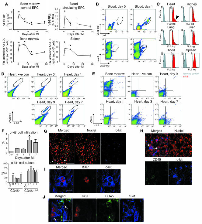
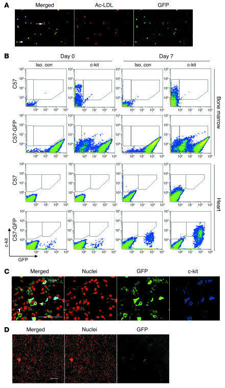
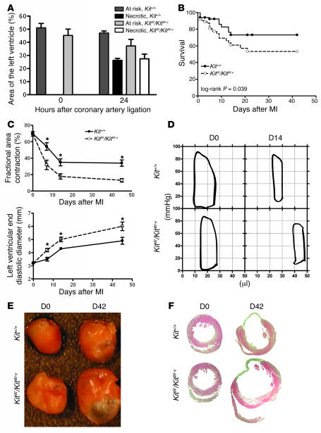
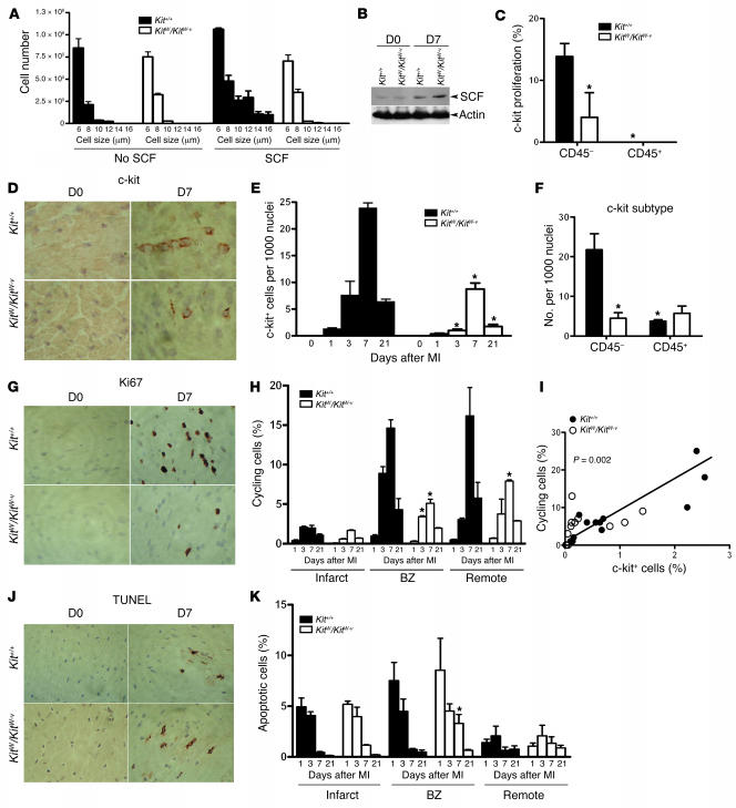
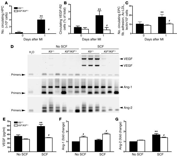
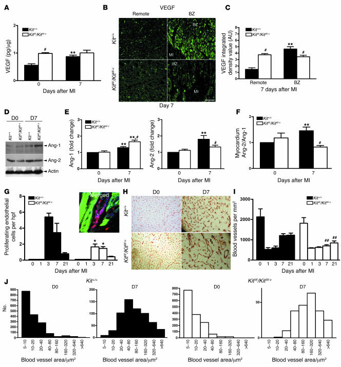
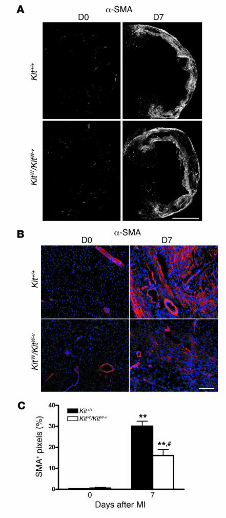
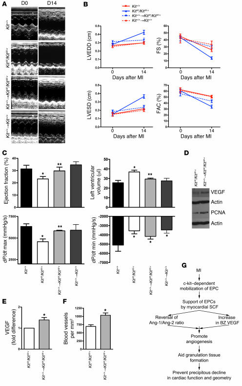
Clinical trials of bone marrow stem/progenitor cell therapy after myocardial infarction (MI) have shown promising results, but the mechanism of benefit is unclear. We examined the nature of endogenous myocardial repair that is dependent on the function of the c-kit receptor, which is expressed on bone marrow stem/progenitor cells and on recently identified cardiac stem cells. MI increased the number of c-kit+ cells in the heart. These cells were traced back to a bone marrow origin, using genetic tagging in bone marrow chimeric mice. The recruited c-kit+ cells established a proangiogenic milieu in the infarct border zone by increasing VEGF and by reversing the cardiac ratio of angiopoietin-1 to angiopoietin-2. These oscillations potentiated endothelial mitogenesis and were associated with the establishment of an extensive myofibroblast-rich repair tissue. Mutations in the c-kit receptor interfered with the mobilization of the cells to the heart, prevented angiogenesis, diminished myofibroblast-rich repair tissue formation, and led to precipitous cardiac failure and death. Replacement of the mutant bone marrow with wild-type cells rescued the cardiomyopathic phenotype. We conclude that, consistent with their documented role in tumorigenesis, bone marrow c-kit+ cells act as key regulators of the angiogenic switch in infarcted myocardium, thereby driving efficient cardiac repair.


Figures

Figure 1
Figure 1. c-kit+ cells increase in infarcted myocardium.
(A) Quantification of EPCs over a time course after MI in wild-type mice. Upperpanels, flow cytometry for VEGFR2+ cells. Lower panels, in vitro splenocyte fibronectin (Fib.) adhesion, acetylated LDL uptake (Ac-LDL), and lectin-binding assay results. Total number of cells was calculated by multiplying percentage of positive cells by total number of cells isolated from both tibia and femur of 1 mouse. n = 3–5 per time point. *P < 0.05 compared to day 0 values. (B) MI causes an increase in VEGFR2+c-kit+Sca-1+ subset of PBMCs. (C) Increase of c-kit+ cells is specific to the injured myocardium (arrowhead). Seven days after MI, c-kit+ cells were not detected in other organs and the peripheral circulation. Gray, isotype control; red, c-kit. (D and E) The c-kit+ cells detected after MI in the heart were homogeneously VEGFR2+ but heterogeneous with respect to CD45 expression. –ve con, negative control. (F) Quantification of the number of c-kit+ cells. Lowerpanel shows that the majority of the c-kit+ cells in the heart were CD45. n = 3 per time point. (GJ) Confocal microscopic images. (G) c-kit+ cells visualized at the infarct border zone both as isolated cells (arrowheads) and in clusters (arrow). Scale bar: 100 μm. (H) The majority of the c-kit+ cells did not express CD45 (arrowhead) although some of the clusters contained CD45-expressing cells (arrow). Scale bar: 50 μm. (I) c-kit+ cells present in the clusters are shown to express Ki67 cell cycling–associated nuclear antigen. Scale bar: 10 μm. (J) c-kit+ cells in the cell cycle (arrowhead) did not coexpress CD45 (arrow). Scale bar: 10 μm.
Figure 2
Figure 2. c-kit+ cells in infarcted myocardium are from the bone marrow.
(A) EPCs from bone marrow chimeric mice carry the GFP transgene. (B) Dual-colored flow cytometry of C57BL/6 (C57) or C57BL/6-GFP bone marrow chimeric mice (C57-GFP) for GFP on the x axis and c-kit on the y axis. In the bone marrow preparation, 74% of c-kit+ cells in the C57-GFP chimeric mice expressed GFP; 74% of c-kit+ cells in the infarcted myocardium in the C57-GFP chimeric mice also expressed GFP. Representative flow cytometry data from 5 independent experiments is shown with results summarized in Table 1. Iso. con, isotype control (C) Confocal micrograph confirming that c-kit+ cells in infarcted myocardium also expressed GFP in chimeric mice (arrow). A GFP+ cell that did not express c-kit (arrowhead) is also visualized in this micrograph. Scale bar: 50 μm. (D) Engraftment of bone marrow–derived cells was minimal when evaluated at 28 days after MI in the bone marrow chimeric mice. Scale bar: 100 μm.
Figure 3
Figure 3. c-kit dysfunction is associated with dilated cardiomyopathy after MI.
(A)Coronary ligation results in a similar volume of myocardium being at risk for infarction and a similar volume becoming necrotic within 24 hours. n =3–4 per group. (B) Actuarial survival is worse in the KitW/KitW–v mice after coronary ligation. n =86 per group. (C) Echocardiography shows rapid decline in cardiac systolic function (fractional area contraction) and rapid ventricular dilation (left ventricular end diastolic diameter in the mutant mice). n =5 per group. *P <0.05. (D) Representative pressure-volume loops of uninfarcted (D0) and infarcted (day 14, D14) Kit+/+ and KitW/KitW–v mice. n =5 per group. Volume is indicated on the x axis and pressure on the y axis. (E) Representative perfusion-fixed hearts before (D0) and 6 weeks after (D42) coronary ligation. Note the ventricular dilation in the KitW/KitW–v mouse. n =5 per group. (F) Representative H&E-stained mid-papillary transverse myocardial sections depicting the pronounced ventricular dilation and larger infarct size in the KitW/KitW–v mice. n =5 per group.
Figure 4
Figure 4. c-kit dysfunction increases apoptosis and decreases mitogenesis.
(A) Incubation of bone marrow cells from Kit+/+ mice with 50 ng/ml of recombinant SCF causes cell proliferation. KitW/KitW–v bone marrow cells had no response to SCF. (B) After coronary ligation, myocardial SCF levels increased in both Kit+/+ and KitW/KitW–v mice. Representative immunoblot of 5 independent experiments is shown. (C) Consistent with the in vitro data, the recruited c-kit+ cells in KitW/KitW–v mice had lower index of proliferation as assessed by Ki67 and c-kit staining and visualized by confocal microscopy of 10 random ×400 fields. n =3 per group. *P <0.05. (D) The total number of c-kit+ cells in KitW/KitW–v mice was lower than in Kit+/+ mice. (E) Quantification of the total number of c-kit–expressing cells in the infarcted myocardium. n =3 per time point per group. (F) The Kit+/+ mice had more CD45 c-kit+ cells than KitW/KitW–v mice. n =3 per group. (G) Nonmyocyte mitogenesis was markedly higher in Kit+/+ mice in the infarct border zone. (H) Quantification of general mitogenesis by region and over a time course. n =3 per time point per group. #P <0.01. BZ, border zone. (I) The number of infiltrating c-kit–expressing cells correlated with the number of cycling cells (r =0.78). (J) Differences in apoptotic cell death as quantified in K were smaller than differences in mitogenesis. n =3 per time point per group. *P <0.05 versus Kit+/+. Magnification, ×200.
Figure 5
Figure 5. c-kit dysfunction is associated with abnormal EPC mobilization and function.
(AC) c-kit function is required for the mobilization of hematopoietic progenitor cells (HPC), VEGFR2+ PBMCs, and EPCs after MI (n =5 per group). **P <0.05 versus Day 0 values; #P < 0.05 versus Kit+/+. (D) RT-PCR reaction for VEGF, angiopoietin-1 (Ang-1), and angiopoietin-2 from bone marrow cells of Kit+/+ or KitW/KitW–v mice cultured for 7 days in the absence or presence of recombinant SCF. SCF led to marked-up regulation of VEGF mRNA only in Kit+/+mice. Angiopoietin-2 levels were higher in Kit+/+ mice. (EG) Quantification of VEGF by ELISA and angiopoietin-2 and angiopoietin-1 levels by immunoblotting and densitometry from cell supernatant described above. SCF caused increased VEGF and higher angiopoietin-2/angiopoietin-1 ratio only in Kit+/+ mice. The values are from 3 independent experiments quantified in triplicate. **P <0.05 versus no SCF values; #P <0.05 versus Kit+/+.
Figure 6
Figure 6. c-kit dysfunction limits myocardial angiogenesis.
(A) VEGF upregulation by total heart ELISA following MI is abrogated in KitW/KitW–v mice. n =5 per group. (B) The VEGF in the KitW/KitW–v mouse myocardium is diffusely present and is not localized to the border zone, as quantified in C. n =4 per group. (C) Integrated density value determined by random sampling in 3 ×400 fields per animal. n =4 per group. (D and E) Kit+/+ responds to MI by increasing the ratio of angiopoietin-2 to angiopoietin-1 whereas KitW/KitW–v responds in the opposite fashion. Representative immunoblot is shown. Data are quantified by immunoblotting and densitometry from 4 independent experiments in triplicates. (F) Angiopoietin-2/angiopoietin-1 ratio. **P <0.05 versus day 0 (D0) values; #P <0.05 versus Kit+/+. (G) Number of endothelial cells (blue is CD31) in the cell cycle (red is Ki67) was quantified using confocal microscopy (actin is green) in 5 random ×400 fields in the border zone. n =3 per group per time point. Number of cycling endothelial cells, which appear magenta in color because of overlap of blue CD31 and red Ki67 staining, was higher in the Kit+/+ mice. *P <0.05. hpf, high-power field. (H) Blood vessel density was assessed by CD31 immunohistochemistry in the border zone. (I) Quantification of the number of CD31+ structures from 5 random ×400 fields converted to mm2 showing diminished angiogenic response in KitW/KitW–v mice. ##P <0.01. (J) Blood vessel size quantification showing the KitW/KitW–v mice vessels to be fewer and of larger caliber. n =5 per group.
Figure 7
Figure 7. c-kit dysfunction limits the formation of repair tissue.
(A) Representative composite images constructed from 15–20 ×10 magnification images demonstrates less α-SMA per infarct area in KitW/KitW–v7 days after MI when compared with Kit+/+mice. (B) Higher magnification images of the border zone and scar area demonstrate that the majority of α-SMA–positive cells are localized in the scar region. (C) Quantitative morphometrical analysis shows a significant difference between the strains. n = 3 per group per time point. **P < 0.05versus day 0 values; #P < 0.01 versus Kit+/+.
Figure 8
Figure 8. Bone marrow rescue also rescues the cardiomyopathic phenotype.
(A) Representative M-mode echocardiographic images in Kit+/+, KitW/KitW–v, and KitW/KitW–v mice whose bone marrow was rescued by Kit+/+ bone marrow after lethal irradiation (Kit+/+KitW/KitW–v) and Kit+/+ mice who received the same dose of irradiation and whose bone marrow was reconstituted from other Kit+/+ mice (Kit+/+Kit+/+), showing the prevention of ventricular dilation in Kit+/+KitW/KitW–v mice. (B) Quantification of echocardiographic parameters: left ventricular end-diastolic diameter (LVEDD), left ventricular end-systolic diameter (LVESD), fractional shortening (FS), and fractional area contraction (FAC). n =5 per group. Bone marrow rescue prevented ventricular dilation and better preserved ventricular systolic function. (C) Invasive hemodynamic measurements 2 weeks after coronary ligation. Bone marrow rescue also rescued parameters of systolic function, such as ejection fraction and dP/dt maximum (max), and prevented ventricular dilation, but did not affect dP/dt minimum (min). n =5–7 per group. *P <0.05 versus Kit+/+; **P <0.05 versus KitW/KitW–v. (D) Bone marrow rescue results in higher myocardial VEGF levels and greater cell cycle activity (PCNA expression). The immunoblots are representative of 4 independent experiments. (E) Quantification of myocardial VEGF from 4 independent experiments performed in triplicate. (F) Bone marrow rescue increases border-zone blood vessels. n =5–7 per group. *P <0.05 versus KitW/KitW–v (E and F).(G) Recruitment of c-kit+ cells from the bone marrow to the injured region of the heart is cardioprotective because it regulates the myocardial balance of angiogenic cytokines.

Comment in

Similar articles

Cited by

References

    1. Orlic D., et al. Mobilized bone marrow cells repair the infarcted heart, improving function and survival. Proc. Natl. Acad. Sci. U. S. A. 2001;98:10344–10349. - PMC - PubMed
    1. Orlic D., et al. Bone marrow cells regenerate infarcted myocardium. Nature. 2001;410:701–705. - PubMed
    1. Fazel S., et al. Current status of cellular therapy for ischemic heart disease. Ann. Thorac. Surg. 2005;79:S2238–S2247. - PubMed
    1. Balsam L.B., et al. Haematopoietic stem cells adopt mature haematopoietic fates in ischaemic myocardium. Nature. 2004;428:668–673. - PubMed
    1. Murry C.E., et al. Haematopoietic stem cells do not transdifferentiate into cardiac myocytes in myocardial infarcts. Nature. 2004;428:664–668. - PubMed

Publication types

MeSH terms

Substances