. 2006 Jul;116(7):2012-21.
doi: 10.1172/JCI27751.

Suppression of canonical Wnt/beta-catenin signaling by nuclear plakoglobin recapitulates phenotype of arrhythmogenic right ventricular cardiomyopathy

Affiliations

Suppression of canonical Wnt/beta-catenin signaling by nuclear plakoglobin recapitulates phenotype of arrhythmogenic right ventricular cardiomyopathy

Eduardo Garcia-Gras et al. J Clin Invest. 2006 Jul.

Abstract

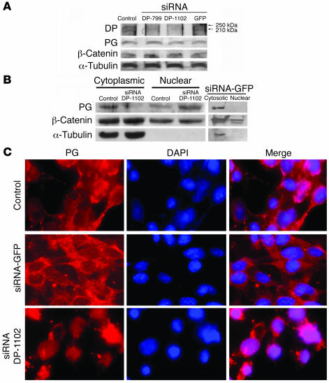
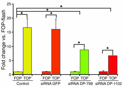
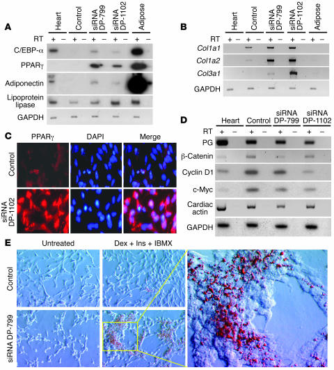
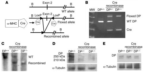
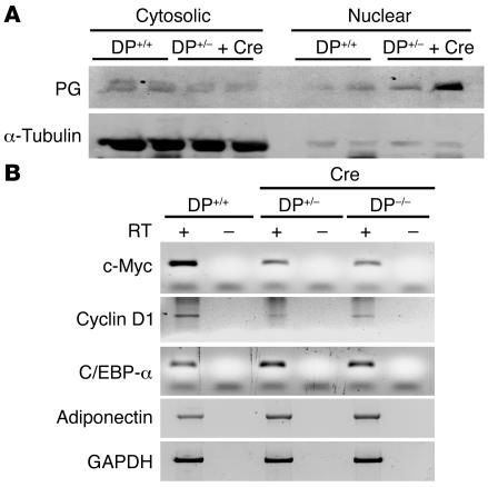
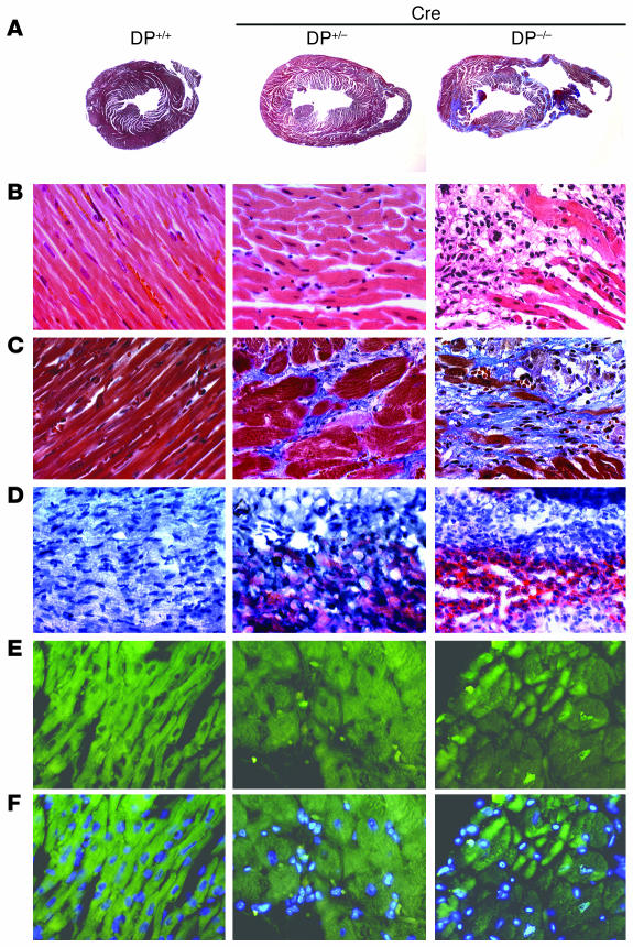
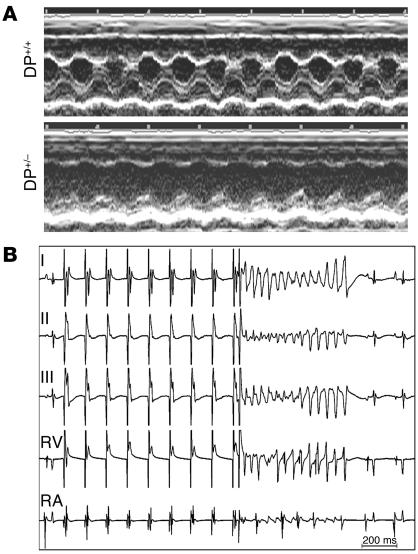
Arrhythmogenic right ventricular dysplasia/cardiomyopathy (ARVC) is a genetic disease caused by mutations in desmosomal proteins. The phenotypic hallmark of ARVC is fibroadipocytic replacement of cardiac myocytes, which is a unique phenotype with a yet-to-be-defined molecular mechanism. We established atrial myocyte cell lines expressing siRNA against desmoplakin (DP), responsible for human ARVC. We show suppression of DP expression leads to nuclear localization of the desmosomal protein plakoglobin and a 2-fold reduction in canonical Wnt/beta-catenin signaling through Tcf/Lef1 transcription factors. The ensuing phenotype is increased expression of adipogenic and fibrogenic genes and accumulation of fat droplets. We further show that cardiac-restricted deletion of Dsp, encoding DP, impairs cardiac morphogenesis and leads to high embryonic lethality in the homozygous state. Heterozygous DP-deficient mice exhibited excess adipocytes and fibrosis in the myocardium, increased myocyte apoptosis, cardiac dysfunction, and ventricular arrhythmias, thus recapitulating the phenotype of human ARVC. We believe our results provide for a novel molecular mechanism for the pathogenesis of ARVC and establish cardiac-restricted DP-deficient mice as a model for human ARVC. These findings could provide for the opportunity to identify new diagnostic markers and therapeutic targets in patients with ARVC.


Figures

Figure 1
Figure 1. Suppression of DP expression in HL-1 cells and nuclear localization of PG.
(A) Immunoblots showing expression levels of DP, PG, β-catenin, and α-tubulin — the latter as a control for loading conditions — in control cells, cells stably transfected with siRNAs against DP, and cells transfected with siRNA against GFP. (B) Immunoblots of subcellular protein extracts probed with an anti-PG antibody. PG was predominantly localized to cytoplasmic protein extracts in control HL-1 cells or cells transfected with siRNA against GFP. In contrast, PG was localized predominantly in the nuclear subfractions in DP-deficient HL-1 cells. (C) Immunofluorescence detection of PG in the nuclei. Shown are cells stained with an anti-PG antibody (left panels), nuclei stained with DAPI (middle panels), and the overlay (right panels) in nontransfected control cells (top panels), cells transfected with siRNA against GFP (middle panels), and DP-deficient HL-1 cells (bottom panels). Magnification, ×400.
Figure 2
Figure 2. TOP-flash assay.
The relative luciferase activity in cells transfected with FOP-flash and TOP-flash vectors in controls cells, cells transfected with siRNA against GFP, and cells transfected with siRNAs against DP are shown. *P < 0.01; Tukey’s test.
Figure 3
Figure 3. Transcriptional switch to adipogenesis and fibrosis and fat droplet accumulation in DP-deficient cells.
(A) Semiquantitative RT-PCR results for C/EBP-α, PPARγ, adiponectin, lipoprotein lipase, and GAPDH, the latter as a control. In addition to control cells and cells transfected with siRNAs, RNA extracts from human heart and adipose tissue are also included. (B) Expression of procollagen genes Col1a1, Col1a2, and Col3a1 in the experimental groups. (C) Immunofluorescence staining of DP-competent and -deficient cells with an anti-PPARγ antibody, showing expression and nuclear localization of PPARγ in DP-deficient cells. (D) Expression levels of mRNAs for selected genes involved in ARVC, including Wnt signaling targets. (E) Accumulation of fat droplets in DP-deficient HL-1 cells treated with dexamethasone, insulin, and 3-isobutyl-1-methylxanthine (Dex + Ins + IBMX). Magnification, ×400.
Figure 4
Figure 4. Targeting vector, genotyping, and detection of expression of DP.
(A) The WT and floxed alleles of Dsp, location of LoxP sequences, sites of BamH1 restriction endonuclease, and location of the probes used in Southern blotting are shown. The construct expressing Cre recombinase under the regulatory control of α-MHC promoter is shown to the left of the panel. (B) Screening of mouse tail DNA by PCR for the presence of floxed and WT alleles and the Cre recombinase transgene in 3 experimental groups. (C) Detection of excised exon 2 and WT alleles by Southern blotting in DNA extracted from the hearts of mice in the experimental groups. (D) Immunoblots on cardiac protein extracts illustrate reduced expression of DP protein in heterozygous mice and its near complete absence in homozygous mice. α-Tubulin was used as a control for loading conditions. (E) Equal expression levels of DP were found in skin tissues from WT, heterozygous, and homozygous DP-deficient mice.
Figure 5
Figure 5. Nuclear localization of PG and transcriptional switch to adipogenesis in DP-deficient mice.
(A) Immunoblots of subcellular protein extracts from WT and DP-deficient mice probed with an anti-PG antibody. PG was predominantly localized to the nuclear protein subfraction in the DP-deficient mice in contrast to WT, which showed predominant localization in cytoplasmic protein subfraction. The difference between cytoplasmic and nuclear PG expression levels in DP+/– mice with α-MHC–Cre mice suggests preferential localization of free (unincorporated) PG to the nucleus. Degradation of the cytoplasmic PG by proteasomes could also contribute to lower levels PG in the cytoplasm. α-Tubulin was used as a control for loading conditions. (B) Detection of expression levels of c-myc and cyclin D1, as target genes for canonical Wnt signaling, and C/EBP-α and adiponectin, as markers of adipogenesis, in WT and DP-deficient mice. Expression levels of c-myc and cyclin D1 were reduced, whereas expression levels of C/EBP-α and adiponectin were increased, in DP-deficient mice compared with WT mice.
Figure 6
Figure 6. Morphological and histological phenotype.
(A) Thin sections of the heart in DP+/+, DP+/–, and DP–/– mice stained with Masson Trichrome, showing ventricular dilatation and fibrosis in DP+/– and DP–/– mice. (B) H&E-stained thin myocardial sections showing myocyte dropout and areas of excess interstitial tissue that comprise cells with adipocytic appearance in the DP–/– mice. (C) Masson Trichrome–stained thin myocardial sections showing extensive areas of fibroadipocytic replacement of myocytes in DP-deficient mice, more prominent in DP–/– mice. (D) Oil Red O–stained thin myocardial sections showing accumulation of fat and adipocytes in DP+/– and DP–/– mice. (E) TUNEL-stained thin myocardial sections showing increased DNA nicking in DP-deficient mice. (F) Same panels as in E overlaid with DAPI-stained nuclei.
Figure 7
Figure 7. Cardiac dysfunction and ventricular arrhythmias.
(A) M-mode section of the left ventricle in DP+/+ and DP+/– mice, showing left ventricular dilatation and reduced fractional shortening in DP+/– mice. (B) Instantaneous ECG and intracardiac electrograms in a heterozygous DP-deficient mouse. The tracings demonstrate an episode of polymorphic ventricular tachycardia induced by a ventricular premature extra stimulus following a ventricular fixed stimulation drive train. RA, right atrial electrogram; RV, right ventricular electrogram. I, II, and III are limb leads I, II, and III on surface ECG.

Comment in

Similar articles

Cited by

References

    1. Corrado D., et al. Spectrum of clinicopathologic manifestations of arrhythmogenic right ventricular cardiomyopathy/dysplasia: a multicenter study. J. Am. Coll. Cardiol. 1997;30:1512–1520. - PubMed
    1. Sen-Chowdhry S., Syrris P., McKenna W.J. Genetics of right ventricular cardiomyopathy. . J. Cardiovasc. Electrophysiol. 2005;16:927–935. - PubMed
    1. Maron B.J., et al. Sudden death in young competitive athletes. Clinical, demographic, and pathological profiles. JAMA. 1996;276:199–204. - PubMed
    1. Tabib A., et al. Circumstances of death and gross and microscopic observations in a series of 200 cases of sudden death associated with arrhythmogenic right ventricular cardiomyopathy and/or dysplasia. Circulation. 2003;108:3000–3005. - PubMed
    1. Kaplan S.R., et al. Structural and molecular pathology of the heart in Carvajal syndrome. . Cardiovasc. Pathol. 2004;13:26–32. - PubMed

Publication types

MeSH terms