. 2006 Jul;11(1):81-92.
doi: 10.1016/j.devcel.2006.04.018.

HIF-dependent hematopoietic factors regulate the development of the embryonic vasculature

Affiliations

HIF-dependent hematopoietic factors regulate the development of the embryonic vasculature

Diana L Ramírez-Bergeron et al. Dev Cell. 2006 Jul.

Abstract

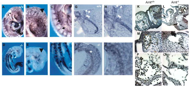
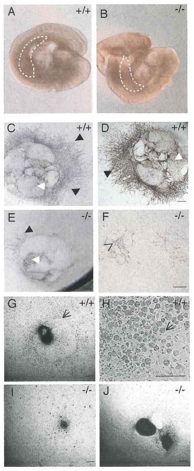
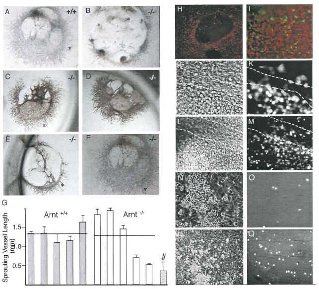
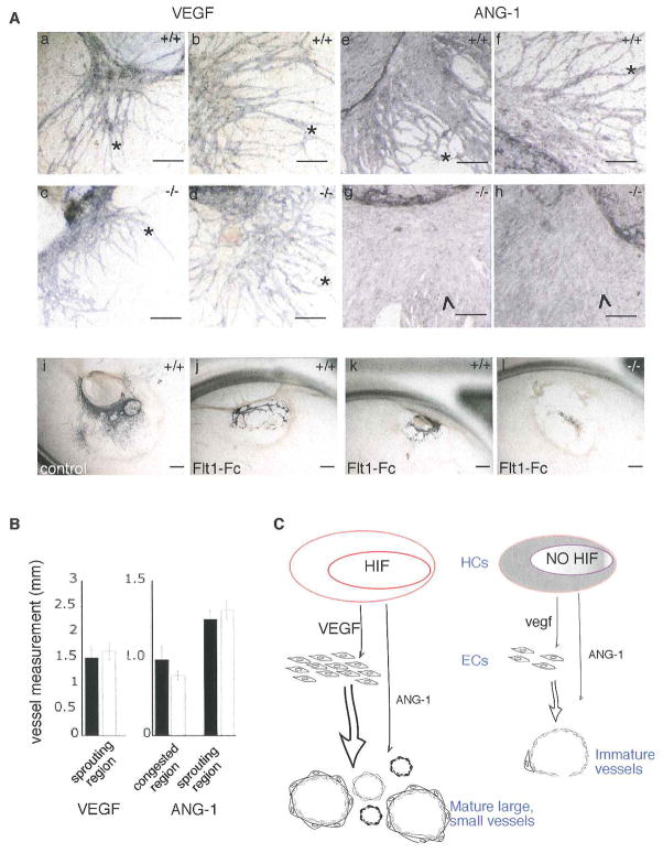
Hypoxia inducible factors (HIFs) regulate adaptive responses to changes in oxygen (O(2)) tension during embryogenesis, tissue ischemia, and tumorigenesis. Because HIF-deficient embryos exhibit a number of developmental defects, the precise role of HIF in early vascular morphogenesis has been uncertain. Using para-aortic splanchnopleural (P-Sp) explant cultures, we show that deletion of the HIF-beta subunit (ARNT) results in defective hematopoiesis and the inhibition of both vasculogenesis and angiogenesis. These defects are rescued upon the addition of wild-type Sca-1(+) hematopoietic cells or recombinant VEGF. Arnt(-/-) embryos exhibit reduced levels of VEGF protein and increased numbers of apoptotic hematopoietic cells. These results suggest that HIF coordinates early endothelial cell emergence and vessel development by promoting hematopoietic cell survival and paracrine growth factor production.


Figures

FIGURE 1
FIGURE 1. Disruption of vascular development in Arnt−/− yolk sacs and embryos
Whole-mount PECAM stained wild type embryo (A,B,C) and yolk sac (G,H), and Arnt−/− embryo (D,E,F) and yolk sac (I,J). The Arnt−/− embryo lacks proper staining of the cardinal vein (A,D arrows) and pharyngeal pouches as seen in the Arnt+/+ embryo (A,D dotted black lines). Compared to wild type, the mutant embryo lacks proper inter- and intra- somitic (black arrows, C and F) and peripheral (arrowheads, B and E) PECAM staining. Striking decreased staining is observed in the AGM region of the Arnt−/− embryo (white dotted lines, A and B). Vascular networks, seen as abnormal branching of the vitelline artery in the yolk sac, are deficient in the Arnt−/− embryo (white arrows, G–J). Immunohistochemical staining with anti-CD34 of Arnt+/+ (K,M,O) and Arnt−/− (L,N,P) 9.5 dpc embryos. The Arnt−/− embryo lacks proper EC staining in the dorsal aorta (M,N, open arrows), and endocardium (O,P, arrows). Enlarged boxed area demonstrates the presence of CD34+ hematopoietic cells in the Arnt−/− embryo (L). Magnifications: A,D, 120X; G,I 150X; B,C,E,F,H,J 200X; K,L 100X; M–P 300X.
FIGURE 2
FIGURE 2. Impaired vascular network formation by Arnt−/− P-Sp explants
P-Sp regions (white dotted lines) from 9.5 dpc Arnt+/+ (A) and Arnt−/− (B) somite matched embryos were removed and cultured on OP9 stromal cells and immunostained with anti-PECAM (C–F). P-Sp explants from Arnt+/+ embryos (C,D), but not Arnt−/− embryos (E,F), formed extensive PECAM+ vascular beds (white arrowheads) and networks (black arrowheads). In contrast, representative Arnt−/− explants exhibit reduced PECAM staining of tissue outgrowth (E) or a few random PECAM+ cells (F, open arrowhead). Wild-type explants (G,H) observed in phase contrast micrographs generated extensive non-adherent round hematopoietic cells (arrows) which are lacking in Arnt−/− P-Sp cultures (I,J). Magnifications: A,B 100X; C,E,G,I, scale bar 100 μm; D,F,J 100X; H, 200X.
FIGURE 3
FIGURE 3. Wild type Sca-1+GFP+ bone marrow cells rescue Arnt−/− P-Sp explants
Sca-1+ cells isolated from the bone marrow of wild type GFP+ mice were added to P-Sp explant cultures and stained for PECAM after 14 days (C–F). As a control, representative untreated Arnt+/+ (A) and Arnt−/− (B) P-Sp cultures are shown. Rescued vascular networks (*) from Arnt−/− explants (C–F) were comparable to wild type controls (A). (G) Measurements of lengths from ten individual vessels extending from the vascular beds expressed as mean ± SEM distance from five independent wild type (grey) and mutant (white) rescued explants. As a control (#) the mean ± SEM from an untreated Arnt−/− culture with minimal vessel staining (B) is shown. The horizontal lines represent average vessel lengths, for the Arnt+/+ and Arnt−/− cultures (P≤ 0.98). (H) Dil-Ac-LDL uptake of endothelial cells from a rescued P-Sp culture showing a peripheral ring of cells that includes the vascular networks. (I) Higher magnification showing no overlap between the DiI-Ac-LDL+ ECs originating from the explant and the GFP+ hematopoietic donor cells in rescued cultures. (J–M) Higher magnification phase contrast (J,L) and counterpart green fluorescent (K,M) micrographs demonstrating track of ECs (dooted white lines) that do not incorporate GFP+ cells. (N–Q) Higher magnification phase contrast (N,P) and green fluorescent (O,Q) micrographs demonstrating nonadherent hematopoietic colonies that are GFP arising from the rescued explants. Magnifications: A–F scale bar 100 μm H, 100X; J–K, 400X; I, L–Q 200X.
FIGURE 4
FIGURE 4
A. VEGF protein levels are reduced in Arnt−/− 9.5 dpc embryos. Transverse sections of Arnt+/+ (a,b,c) and Arnt−/− (d,e,f) embryos probed for Vegf(b,e) and Pgk (c,f) showing no overall differences in expression. As a comparison, hematoxylin stained Arnt+/+ (a) and Arnt−/− (d) serial sections are shown. VEGF immunohistochemistry of transverse sections of 9.5 dpc (28 somites) Arnt+/+ (g) and Arnt−/− (h) embryos showing reduced protein expression in the mutant embryo. Expression is particularly reduced in the hematopoietic compartment (higher magnification inlay) of the Arnt−/− embryo (arrow). Dorsal aorta (da); hematopoietic cells (HCS); heart (ht); neural tube (nt). (i) VEGF levels from whole cell or postnuclear lysates of individual 9.5 dpc (21 somites) Arnt+/+ (black) and Arnt−/− (white) embryos analyzed by ELISA. Error bars indicate standard error of the mean (n=3, (*) P<0.005). B. Increased apoptosis in Arnt−/− embryos. TUNEL immunostaining (FITC, green) of transverse sections counterstained with hematoxylin (a,b) or nuclear DAPI (blue) (c,d), and hematoxylin stained sagittal sections (e,f) of Arnt+/+ (a,c,e) and Arnt−/− (b,d,f) embryos. An increase in TUNEL+ HCs was detected in the dorsal aorta (d, arrowheads) of Arnt−/− transverse sections compared to the modest TUNEL staining observed in the Arnt+/+ control (c). Increased numbers of TUNEL+ cells in the dorsal aorta (f, arrows) and intrasomitic (f, dotted lines) regions are observed in Arnt−/− sagittal sections. Comparison of apoptotic cells in the dorsal aortic region of Arnt+/+ (black) and Arnt−/− (white) transverse sections (g). Numbers of apoptotic cells are expressed as total numbers of FITC+ cells (*) P<0.005. Magnifications: 50X A a–f, Ba,b,e,f; 100X A g–h, B c,d (inserts 400X).
Figure 5
Figure 5
A. Treatment of P-Sp explants with VEGF or Ang-1 promotes vessel growth Addition of 100 ng/ml VEGF to Arnt−/− cultures (c,d) promotes fine patterning and branching in the vascular networks (*). Arnt+/+ cultures (a,b) are shown as controls. Addition of 300 ng/ml Ang-1 does not result in proper angiogenesis but promotes congestion (A) in the vascular networks in Arnt−/− cultures (g,h) as well as the Arnt+/+ P-Sps (e,f; P<0.722) However, the congestion was worse in the Arnt−/− explants. Addition of 300 ng/ml of Flt-1-Fc (j–l) inhibited proper vessel development as compared to control (i) P-Sp explants. All explants were stained for PECAM. Magnifications: scale bars 100 μm. B. Measurement of vessel areas from VEGF or Ang-1 treated P-Sp explants. Ten sprouting vessel measurements from five independent Arnt−/− and Arnt+/+ P-Sp explants were analyzed. Results are shown as mean vessel lengths (±SEM) for wild type (black) and mutant (white) VEGF treated or Ang-1 treated explants. Note that Ang-1 promoted vessel congestion. C. Proper vessel development requires HIF- dependent factors supplied by hematopoietic cells. Differentiation of hematopoietic cells and vasculature is HIF-dependent during three separate events. (1) HIF promotes the survival of hematopoietic cells. (2) Hematopoietic cells serve as a paracrine source of VEGF which induces vasculogenesis by increasing the production and proliferation of endothelial cells. (3) Hematopoietic cells are also a source of Ang-1, promoting angiogenesis, as measured by the remodeling and maturation of vessels (Takakura et al., 2000). Loss of Arnt results in HIF deficiency, increased numbers of apoptotic cells in the hematopoietic compartment, and a reduction of VEGF production.

Similar articles

Cited by

References

    1. Adelman DM, Gertsenstein M, Nagy A, Simon MC, Maltepe E. Placental cell fates are regulated in vivo by HIF-mediated hypoxia responses. Genes Dev. 2000;14:3191–3203. - PMC - PubMed
    1. Adelman DM, Maltepe E, Simon MC. Multilineage embryonic hematopoiesis requires hypoxic ARNT activity. Genes Dev. 1999;13:2478–2483. - PMC - PubMed
    1. Arai P, Hirao A, Suda T. Regulation of hematopoietic stem cells by the niche. Trends Cardiovasc Med. 2005;15:75–79. - PubMed
    1. Brusselmans K, Bono F, Collen D, Herbert JM, Carmeliet P, Dewerchin M. A novel role for vascular endothelial growth factor as an autocrine survival factor for embryonic stem cells during hypoxia. J Biol Chem. 2005;280:3493–3499. - PubMed
    1. Carmeliet P. Angiogenesis in life, disease and medicine. Nature. 2005;438:932–936. - PubMed

Publication types

MeSH terms

Substances