. 2006 Sep 15;575(Pt 3):845-54.
doi: 10.1113/jphysiol.2006.111252. Epub 2006 Jul 6.

The inotropic effect of cardioactive glycosides in ventricular myocytes requires Na+-Ca2+ exchanger function

Affiliations

The inotropic effect of cardioactive glycosides in ventricular myocytes requires Na+-Ca2+ exchanger function

Julio Altamirano et al. J Physiol. .

Abstract

Glycoside-induced cardiac inotropy has traditionally been attributed to direct Na(+)-K(+)-ATPase inhibition, causing increased intracellular [Na(+)] and consequent Ca(2+) gain via the Na(+)-Ca(2+) exchanger (NCX). However, recent studies suggested alternative mechanisms of glycoside-induced inotropy: (1) direct activation of sarcoplasmic reticulum Ca(2+) release channels (ryanodine receptors; RyRs); (2) increased Ca(2+) selectivity of Na(+) channels (slip-mode conductance); and (3) other signal transduction pathways. None of these proposed mechanisms requires NCX or an altered [Na(+)] gradient. Here we tested the ability of ouabain (OUA, 3 microm), digoxin (DIG, 20 microm) or acetylstrophanthidin (ACS, 4 microm) to alter Ca(2+) transients in completely Na(+)-free conditions in intact ferret and cat ventricular myocytes. We also tested whether OUA directly activates RyRs in permeabilized cat myocytes (measuring Ca(2+) sparks by confocal microscopy). In intact ferret myocytes (stimulated at 0.2 Hz), DIG and ACS enhanced Ca(2+) transients and cell shortening during twitches, as expected. However, prior depletion of [Na(+)](i) (in Na(+)-free, Ca(2+)-free solution) and in Na(+)-free solution (replaced by Li(+)) the inotropic effects of DIG and ACS were completely prevented. In voltage-clamped cat myocytes, OUA increased Ca(2+) transients by 48 +/- 4% but OUA had no effect in Na(+)-depleted cells (replaced by N-methyl-d-glucamine). In permeabilized cat myocytes, OUA did not change Ca(2+) spark frequency, amplitude or spatial spread (although spark duration was slightly prolonged). We conclude that the acute inotropic effects of DIG, ACS and OUA (and the effects on RyRs) depend on the presence of Na(+) and a functional NCX in ferret and cat myocytes (rather than alternate Na(+)-independent mechanisms).


Figures

Figure 1
Figure 1. Protocol for assessing inotropy under Na+-free conditions in ferret ventricular myocytes
The cell was field stimulated at a relatively low frequency (0.2 Hz). Shortening was measured continually using a video edge-detector system. Upon steady state, the external NT was replaced by a Na+-free, Ca2+-free solution. The cell was kept in this solution for about 10 min until the cytosol was depleted of Na+. Then, normal external [Ca2+] (2 mm) was restored to the bath and electrical stimulation proceeded until a new steady state in shortening and [Ca2+]i transients (not shown) was reached. ACS (4 μm) or DIG (20 μm) was then applied in Na+-free conditions to evaluate inotropy. Inset shows the normal inotropic effect of 4 μm ACS on cell shortening in the presence of external Na+.
Figure 2
Figure 2. Pooled data of the effect of 4 μm acetylstrophanthidin (ACS, A) and 20 μm digoxin (DIG, B) on shortening in ferret ventricular myocytes
Data were measured at the indicated time after exposure to ACS or DIG in the presence or absence of Na+. Each data point represents the average of six cells from six different animals for ACS, and four cells from three different animals for DIG. Shortening is expressed as percentage change normalized to the recordings before addition of glycoside. *P < 0.05 compared with control.
Figure 3
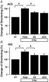
Figure 3. Changes in shortening of ferret ventricular myocytes exposed to Na+-free solution, before and after addition of 4 μm acetylstrophanthidin (ACS, A) and 20 μm digoxin (DIG, B)
Data were normalized to shortening measured in NT. Each bar represents the pooled data for six and four different cells exposed to ACS and DIG, respectively. Peak shortening was measured in [Na+]i depleted cells (as illustrated in Fig. 1) 2–5 min after external Ca2+ had been readmitted. This transient increase in shortening probably represents the removal of remaining intracellular Na+, with the consequent increase in Ca2+. Steady-state (SS) shortening was measured ∼20 min after external Ca2+ readmission. ACS and DIG data were measured 20 min after glycoside exposure in Na+-free, 2 mm Ca2+ solution.
Figure 4
Figure 4. Ca2+ transients in voltage-clamped cat ventricular cells exposed to 3 μm ouabain (OUA) in control (A and C) and Na+-free solution (B and D)
Cells were depolarized to +10 mV (from a holding potential of −70 mV) at 0.5 Hz. A, shows the expected increase in [Ca2+]i in a representative cell in the presence of external Na+. B, cells were incubated previously in a Na+-free solution until [Na+]i had been depleted and then exposed to OUA in a Na+-free, 1 mm Ca2+ solution. [Ca2+]i transients were not affected by exposure to OUA in a Na+-free solution. However, rapid increase in external [Ca2+] to 5 mm (inset) or the β adrenergic agonist isoprenaline (1 μm, not shown) could still substantially increase the amplitude of the [Ca2+]i transient in Na+-free conditions. C and D, pooled data for the normalized Ca2+ transients in NT and Na+-free solutions, respectively (numbers in parentheses indicate numbers of cells studied from two and three animals for control and Na+-free experiments, respectively). There was no significant increase in the amplitude of the Ca2+ transient in Na+-free solutions, whereas in NT it significantly increased (*P < 0.05 compared with control).
Figure 5
Figure 5. Effect of ouabain (OUA, 3 μm) on resting [Ca2+]i sparks in streptolysin-O-permeabilized cat ventricular myocytes
A, spontaneous sparks measured with line-scan confocal microscopy in a Na+-free, 50 nm Ca2+ solution before and 5 min after exposure to OUA. B, shows the time course of the pooled data from nine cells from three animals for the resting [Ca2+]i spark frequency (CaSpF) before and during exposure to OUA. C, illustrates typical spontaneous [Ca2+]i sparks before and during OUA exposure. Both [Ca2+]i sparks were measured in the same cell and at the same site in order to avoid out of focus problems and cell–cell variability in [Ca2+]i sparks. There was a significant increase in spark duration (FDHM, P < 0.05). However, neither Ca2+ spark amplitude nor spatial spread (FWHM) was significantly affected (see Table 1).

References

    1. Arnon A, Hamlyn JM, Blaustein MP. Ouabain augments Ca2+ transients in arterial smooth muscle without raising cytosolic Na+ Am J Physiol Heart Circ Physiol. 2000;279:H679–H691. - PubMed
    1. Bassani JW, Bassani RA, Bers DM. Relaxation in rabbit and rat cardiac cells: species-dependent differences in cellular mechanisms. J Physiol. 1994a;476:279–293. - PMC - PubMed
    1. Bassani RA, Bassani JW, Bers DM. Relaxation in ferret ventricular myocytes: unusual interplay among calcium transport systems. J Physiol. 1994b;476:295–308. - PMC - PubMed
    1. Bassani RA, Bassani JW, Bers DM. Relaxation in ferret ventricular myocytes: role of the sarcolemmal Ca2+ ATPase. Pflugers Arch. 1995;430:573–578. - PubMed
    1. Bers DM. Excitation-Contraction Coupling and Cardiac Contractile Force. 2. Netherlands: Kluwer Academic Publishers; 2001.

Publication types

MeSH terms

LinkOut - more resources