. 2006 Jul;44(7):2595-600.
doi: 10.1128/JCM.00144-06.

Use of in vitro assays to determine effects of human serum on biological characteristics of Acanthamoeba castellanii

Affiliations

Use of in vitro assays to determine effects of human serum on biological characteristics of Acanthamoeba castellanii

James Sissons et al. J Clin Microbiol. 2006 Jul.

Abstract

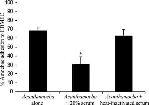
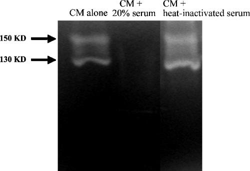
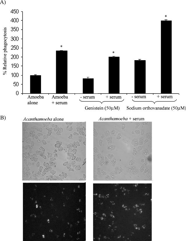
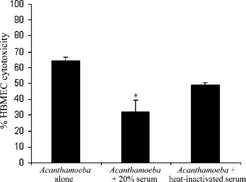
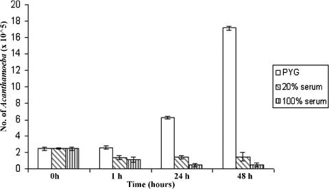
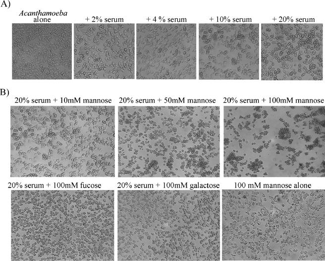
Normal human serum inhibits Acanthamoeba (encephalitis isolate) binding to and cytotoxicity of human brain microvascular endothelial cells, which constitute the blood-brain barrier. Zymographic assays revealed that serum inhibits extracellular protease activities of acanthamoebae. But it is most likely that inhibition of specific properties of acanthamoebae is a consequence of the initial amoebicidal-amoebistatic effects induced by serum. For example, serum exhibited amoebicidal effects; i.e., up to 50% of the exposed trophozoites were killed. The residual subpopulation, although viable, remained static over longer incubations. Interestingly, serum enhanced the phagocytic ability of acanthamoebae, as measured by bacterial uptake. Overall, our results demonstrate that human serum has inhibitory effects on Acanthamoeba growth and viability, protease secretions, and binding to and subsequent cytotoxicity for brain microvascular endothelial cells. Conversely, Acanthamoeba phagocytosis was stimulated by serum.


Figures

FIG. 1.
FIG. 1.
Serum significantly (*, P < 0.05) inhibited Acanthamoeba adhesion to HBMEC. These results are representative of three independent experiments performed in triplicate. Bars represent standard errors.
FIG. 2.
FIG. 2.
Serum inhibits extracellular proteases of acanthamoebae. CM was collected, and proteolytic activities were determined by zymographic assays. Note that serum abolished Acanthamoeba proteases. These results are representative of three independent experiments. KD, kilodaltons.
FIG. 3.
FIG. 3.
(A) Serum enhances Acanthamoeba phagocytosis with live E. coli K-12. Untreated acanthamoebae were used as 100% phagocytosis, and levels of phagocytosis in serum and/or inhibitor-treated acanthamoebae are shown as percent change. Serum exhibits a significant (*, P < 0.05) increase in Acanthamoeba phagocytosis. These results are representative of three independent experiments performed in triplicate. Bars represent standard errors. (B) Representative effects of serum on Acanthamoeba uptake of heat-killed, FITC-labeled E. coli. Note that serum enhanced Acanthamoeba uptake of FITC-labeled E. coli. Magnification, ×200.
FIG. 4.
FIG. 4.
Serum significantly (*, P < 0.05) inhibited Acanthamoeba-mediated HBMEC cytotoxicity. Heat inactivation partially restored Acanthamoeba-mediated HBMEC cytotoxicity (not significant). These results are representative of three independent experiments performed in triplicate. Bars represent standard errors.
FIG. 5.
FIG. 5.
Human serum exhibits amoebicidal and amoebistatic effects. At a concentration of 20%, serum exhibited an initial amoebicidal effects, followed by an amoebistatic effect. In contrast, 100% serum exhibited an amoebicidal effect for up to 24 h, followed by an amoebistatic effect. These results are representative of three independent experiments performed in triplicate. Bars represent standard errors.
FIG. 6.
FIG. 6.
(A) Serum induced Acanthamoeba clustering in a concentration-dependent manner. (B) Mannose, but not other sugars, enhanced serum-induced clustering of acanthamoebae in a concentration-dependent manner. These results are representative of three independent experiments.

References

    1. Alsam, S., K. S. Kim, M. Stins, A. O. Rivas, J. Sissons, and N. A. Khan. 2003. Acanthamoeba interactions with human brain microvascular endothelial cells. Microb. Pathog. 35(6):235-241. - PubMed
    1. Alsam, S., J. J. Sissons, S. Jayasekera, and N. A. Khan. 2005. Extracellular proteases of Acanthamoeba castellanii (encephalitis isolate belonging to T1 genotype) contribute to increased permeability in an in vitro model of the human blood-brain barrier. J. Infect. 51(2):150-156. - PubMed
    1. Alsam, S., J. Sissons, R. Dudley, and N. A. Khan. 2005. Mechanisms associated with Acanthamoeba castellanii (T4) phagocytosis. Parasitol. Res. 96(6):402-409. - PubMed
    1. Ferrante, A., and B. Rowan-Kelly. 1983. Activation of the alternative pathway of complement by Acanthamoeba culbertsoni. Clin. Exp. Immunol. 54(2):477-485. - PMC - PubMed
    1. Ferrante, A. 1991. Immunity to Acanthamoeba. Rev. Infect. Dis. 13:S403-S409. - PubMed

Publication types

MeSH terms

LinkOut - more resources