Prion-induced amyloid heart disease with high blood infectivity in transgenic mice
- PMID: 16825571
- PMCID: PMC1820586
- DOI: 10.1126/science.1128635
Prion-induced amyloid heart disease with high blood infectivity in transgenic mice
Abstract
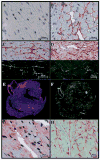
We investigated extraneural manifestations in scrapie-infected transgenic mice expressing prion protein lacking the glycophosphatydylinositol membrane anchor. In the brain, blood, and heart, both abnormal protease-resistant prion protein (PrPres) and prion infectivity were readily detected by immunoblot and by inoculation into nontransgenic recipients. The titer of infectious scrapie in blood plasma exceeded 10(7) 50% infectious doses per milliliter. The hearts of these transgenic mice contained PrPres-positive amyloid deposits that led to myocardial stiffness and cardiac disease.
Figures


Similar articles
-
Anchorless prion protein results in infectious amyloid disease without clinical scrapie.Science. 2005 Jun 3;308(5727):1435-9. doi: 10.1126/science.1110837. Science. 2005. PMID: 15933194
-
Ultrastructure and pathology of prion protein amyloid accumulation and cellular damage in extraneural tissues of scrapie-infected transgenic mice expressing anchorless prion protein.Prion. 2017 Jul 4;11(4):234-248. doi: 10.1080/19336896.2017.1336274. Epub 2017 Jul 31. Prion. 2017. PMID: 28759310 Free PMC article.
-
Fatal transmissible amyloid encephalopathy: a new type of prion disease associated with lack of prion protein membrane anchoring.PLoS Pathog. 2010 Mar 5;6(3):e1000800. doi: 10.1371/journal.ppat.1000800. PLoS Pathog. 2010. PMID: 20221436 Free PMC article.
-
Is the prion structure solved?Arch Immunol Ther Exp (Warsz). 1997;45(2-3):121-40. Arch Immunol Ther Exp (Warsz). 1997. PMID: 9597078 Review.
-
The neuropathological phenotype in transgenic mice expressing different prion protein constructs.Philos Trans R Soc Lond B Biol Sci. 1994 Mar 29;343(1306):415-23. doi: 10.1098/rstb.1994.0038. Philos Trans R Soc Lond B Biol Sci. 1994. PMID: 7913760 Review.
Cited by
-
Defining the conformational features of anchorless, poorly neuroinvasive prions.PLoS Pathog. 2013;9(4):e1003280. doi: 10.1371/journal.ppat.1003280. Epub 2013 Apr 18. PLoS Pathog. 2013. PMID: 23637596 Free PMC article.
-
Shedding light on prion disease.Prion. 2015;9(4):244-56. doi: 10.1080/19336896.2015.1065371. Prion. 2015. PMID: 26186508 Free PMC article.
-
Crucial role for prion protein membrane anchoring in the neuroinvasion and neural spread of prion infection.J Virol. 2011 Feb;85(4):1484-94. doi: 10.1128/JVI.02167-10. Epub 2010 Dec 1. J Virol. 2011. PMID: 21123371 Free PMC article.
-
Unusual cerebral vascular prion protein amyloid distribution in scrapie-infected transgenic mice expressing anchorless prion protein.Acta Neuropathol Commun. 2013 Jun 19;1:25. doi: 10.1186/2051-5960-1-25. Acta Neuropathol Commun. 2013. PMID: 24252347 Free PMC article.
-
Detection of prion infectivity in fat tissues of scrapie-infected mice.PLoS Pathog. 2008 Dec;4(12):e1000232. doi: 10.1371/journal.ppat.1000232. Epub 2008 Dec 5. PLoS Pathog. 2008. PMID: 19057664 Free PMC article.
References
Publication types
MeSH terms
Substances
Grants and funding
LinkOut - more resources
Full Text Sources
Other Literature Sources
Medical
Molecular Biology Databases

